. 2020 Nov 18;21(22):8709.
doi: 10.3390/ijms21228709.

Discovery and Design of Novel Small Molecule GSK-3 Inhibitors Targeting the Substrate Binding Site

Affiliations

Discovery and Design of Novel Small Molecule GSK-3 Inhibitors Targeting the Substrate Binding Site

Ido Rippin et al. Int J Mol Sci. .

Abstract

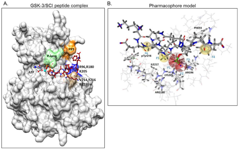
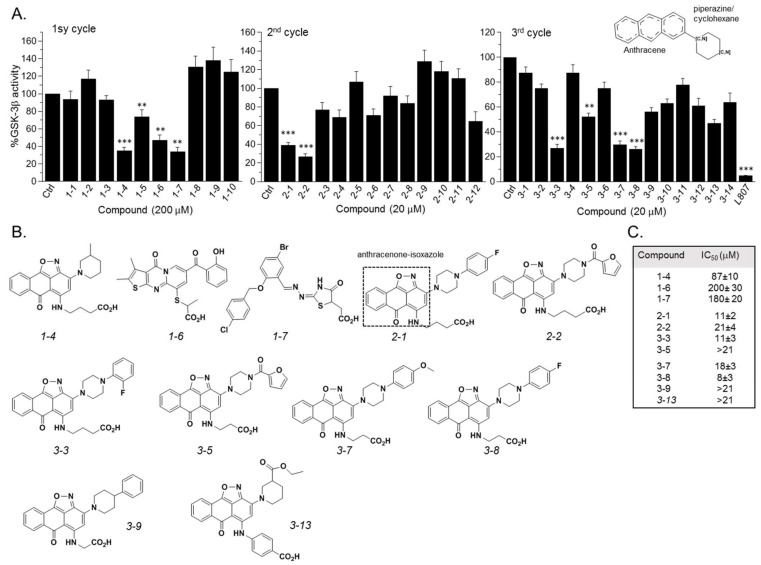
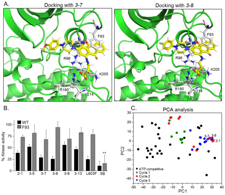
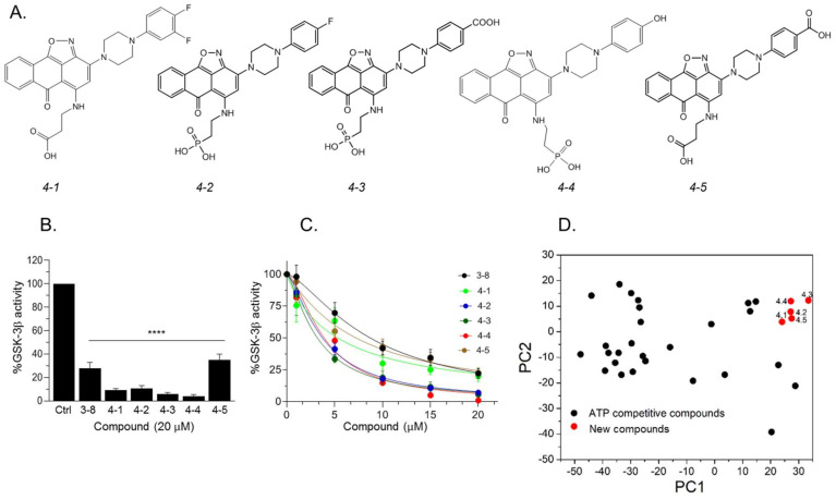
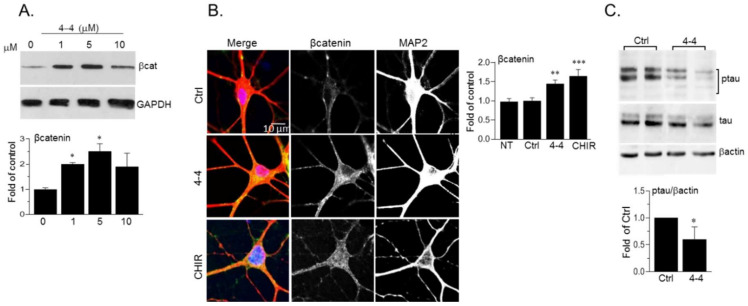
The serine/threonine kinase, GSK-3, is a promising drug discovery target for treating multiple pathological disorders. Most GSK-3 inhibitors that were developed function as ATP competitive inhibitors, with typical limitations in specificity, safety and drug-induced resistance. In contrast, substrate competitive inhibitors (SCIs), are considered highly selective, and more suitable for clinical practice. The development of SCIs has been largely neglected in the past because the ambiguous, undefined nature of the substrate-binding site makes them difficult to design. In this study, we used our previously described structural models of GSK-3 bound to SCI peptides, to design a pharmacophore model and to virtually screen the "drug-like" Zinc database (~6.3 million compounds). We identified leading hits that interact with critical binding elements in the GSK-3 substrate binding site and are chemically distinct from known GSK-3 inhibitors. Accordingly, novel GSK-3 SCI compounds were designed and synthesized with IC50 values of~1-4 μM. Biological activity of the SCI compound was confirmed in cells and in primary neurons that showed increased β-catenin levels and reduced tau phosphorylation in response to compound treatment. We have generated a new type of small molecule GSK-3 inhibitors and propose to use this strategy to further develop SCIs for other protein kinases.

Keywords: GSK-3; peptides; pharmacophore; small molecules; substrate competitive inhibitors; virtual screening.


Conflict of interest statement

The authors declare no conflict of interest.

Figures

Figure 1
Figure 1
Design of a pharmacophore model based on GSK-3β binding model with substrate competitive inhibitor (SCI) peptide (A) Structural model of GSK-3β bound to our previously described SCI peptide L803F. The primed. phosphate S10(p) in the peptide interacts with the phosphate-binding pocket (Arg 96, Arg 180, Ly 205, marked blue), Phe12, at the C-terminal end of the peptide, interacts with Phe93 located in the “89–95” loop (orange), Ala3 in the peptide interacts with pTyr216, and Pro5 in the peptide interacts with Val 214 and Ile217, all residues that form the “hydrophobic patch” (beige). The P-loop including Phe67 is in green, the ATP molecule and Mg+2 (cyan balls) are shown. (B) The pharmacophore model is composed of two hydrogen bond acceptor features (F1, F2: red vectors), one anionic feature (F3: red porcupine shape), and three hydrophobic features (F4, F5, F6: yellow spheres). Excluded volumes were placed in positions that are sterically claimed by the protein environment. GSK-3 interacting residues are marked.
Figure 2
Figure 2
Discovery of GSK-3 SCI hits. (A) In vitro GSK-3β kinase assays were conducted with selected hits identified in the three iterative virtual screening cycles. Structure of compounds is summarized in Tables S1–S3. Ctrl represents GSK-3 activity no compounds (100%). Results are mean of three independent experiments ± SEM using one-way ANOVA with Dunnett’s post hoc test. ** p < 0.01 *** p < 0.001. The anthracene core used in the third search cycle is illustrated at the top right panel. The SCI peptide L807mts (L807) was used as a reference in the 3rd cycle assays. (B) Chemical structures of selected best hits of each cycle. The anthracenone–isoxazole core is highlighted in compound 2-1. (C) IC50 values of selected best hits.
Figure 3
Figure 3
GSK-3 SCI compounds interact with the kinase substrate binding site and are chemically unique. (A) Docking models of compounds 3-7 and 3-8 with GSK-3β. Key interacting GSK-3β residues are highlighted, and detailed interactions are summarized in Table 1. The carboxylic acid moiety form salt bridges and H-bonding with the GSK-3-phosphate-binding pocket, Arg 96, Arg 180, Lys 205, and with Val 214. The anthracenone–isoxazole core forms π–π stacking interactions with Phe 93. Interactions were analyzed by the Maestro software. (B) In vitro kinase assays were performed with GSK-3β (WT) or with the F93A mutant in the presence of indicated compounds, L803F (20 μM each), and SB216763 (1 μM). Results are mean of three independent experiments ± SEM analyzed by one way ANOVA with Dunnett’s multiple comparisons. ** p < 0.01 with inhibitor vs no inhibitor (C) Principal component analysis (PCA) analysis of all compounds discovered through the three search cycles together with representative GSK-3-ATP competitive inhibitors (listed in Table S4). The first and second PCs accounted for 81.8% and 7.7% of the original variance and are shown at the X- and Y-axis respectively. Black circles represent the ATP competitive inhibitors, colored circles represent compounds from cycle 1 (green), 2 (red) and 3 (blue). Circles with yellow dots represent compounds 2-1, 3-7 and 3-8.
Figure 4
Figure 4
Newly designed GSK-3 SCI molecules. (A) Structures of new molecules 4-1 to 4-5. (B) In vitro GSK-3β kinase assays were conducted with new compounds and 3-8 (20 μM each). Results show the percentage of GSK-3β activity without inhibitor (100%) and represent the mean of three independent experiments ± SEM analyzed by one way ANOVA with Dunnett’s multiple comparisons. **** p < 0.0001. (C) Dose–response curves of GSK-3β inhibition of new compounds and as compared to 3-8. Results are mean of three independent experiments ± SEM. For 4-3 and 4-4 * p < 0.05 for all concentrations, for the rest of the molecules, * p < 0.05 at concentrations ≥ 5 μM as determined by one way ANOVA with Dunnett’s multiple comparisons, new compounds vs. 3-8. (D) PCA analysis of new compounds 4-1 to 4-5 together with GSK-3-ATP competitive inhibitors (listed in Table S4). The first and second PCs accounted for 81.8% and 7.7% of the original variance and are shown at the X- and Y-axis respectively. Black circles represent the ATP competitive inhibitors, red circles represent new compounds.
Figure 5
Figure 5
Docking new GSK-3 SCI molecules in GSK-3 substrate binding site. Docking models of GSK-3β bound to 4-2, 4-3 and 4-4. Detailed interactions are also summarized in Table 2. Like 3-8 and the other leading hits, the new molecules interacted with the GSK-3 phosphate-binding pocket, Phe 93 and Val 214. 4-3 and 4-4 showed additional interactions with Phe 67, Ly 85 (4-3), or, Ser 66 (4-4). Interactions were analyzed by the Maestro software.
Figure 6
Figure 6
Kinetic analysis of new compounds. (A) Michaelis Menten plots of ATP competitive assays with 4-3 and 4-4. The corresponding Lineaver–Burk plots are shown at the right panel. (B) Michaelis Menten plots of substrate competitive assays with 4-3 and 4-4. The corresponding Lineaver–Burk plots are shown at the right panel. (C) In vitro kinase assays of F93A mutant in the presence of 4-1 through 4-5 (20 μM each). Results are mean of three independent experiments ± SEM using one-way ANOVA with Dunnett’s post hoc test. (D) GSK-3α kinase assays were performed with 4-1 through 4-5 (20 μM each). Results the mean of three independent experiments ± SEM using one-way ANOVA with Dunnett’s post hoc test. **** p < 0.0001 treated vs. control. (E) GSK-3β kinase assays were performed with 4-3 and 4-4 (20 μM) in the presence of 0.05% Triton × 100. (F) Kinase assays were performed with a representative repertoire of protein kinases in the presence of 4-4 (10 μM). Inhibition by 4-4 is presented as a percentage of kinase activity in the control assay with no inhibitor. Results are means ± SD of two independent experiments performed in duplicates. GSK-3α/β are marked red.
Figure 7
Figure 7
Biological activity of 4-4. (A) SH-SY5Y cells were treated with 4-4 for 4 h and levels of cytoplasmic β-catenin were determined by immunoblot analysis. Densitometry analysis is shown on the lower panel. Results are mean of three independent experiments ± SEM * p < 0.05 by one-way ANOVA with Dunnett’s post hoc test (B) Hippocampal mouse neurons were treated with 4-4 (5 μM), or CHIR99021 (CHIR, 10 μM) for 4 h. Cells were co-stained with anti-β-catenin and anti-MAP2 antibodies. Images show overlapping β-catenin (green) and MAP2 (red) staining along with respective split channels (shown in grey). The β-catenin signal was evaluated by Image J Colocalization-finder plugin. Results present the mean of 60 cells ± SEM, ** p < 0.01, *** p < 0.0001 by one-way ANOVA with Dunnett’s post hoc test. NT- non treated. (C) Hippocampal mouse neurons were treated with 4-4 (20 μM) for 4 h. Levels of phosphorylated tau (Ser 396), tau, and β-actin were determined by immunoblot analysis. Bar graph represents densitometry analysis of ptau/βactin and results are mean of three independent experiments ± SEM using Student’s t-test. * p < 0.05. For all panels, Ctrl or 0 concentration represents cells treated with vehicle (DMSO/1%Tween 80 at matched dilutions—1:2000–4000).

Similar articles

Cited by

References

    1. Manning G., Whyte D.B., Martinez R., Hunter T., Sudarsanam S. The protein kinase complement of the human genome. Science. 2002;298:1912–1934. doi: 10.1126/science.1075762. - DOI - PubMed
    1. Hanks S.K., Quinn A.M., Hunter T. The protein kinase family: Conserved features and deduced phylogeny of the catalytic domains. Science. 1988;241:42–52. doi: 10.1126/science.3291115. - DOI - PubMed
    1. Taylor S.S., Radzio-Andzelm E., Hunter T. How do protein kinases discriminate between serine/threonine and tyrosine? Structural insights from the insulin receptor protein-tyrosine kinase. FASEB J. 1995;9:1255–1266. doi: 10.1096/fasebj.9.13.7557015. - DOI - PubMed
    1. Bain J., Plater L., Elliott M., Shpiro N., Hastie C.J., McLauchlan H., Klevernic I., Arthur J.S., Alessi D.R., Cohen P. The selectivity of protein kinase inhibitors: A further update. Biochem. J. 2007;408:297–315. doi: 10.1042/BJ20070797. - DOI - PMC - PubMed
    1. Davis M.I., Hunt J.P., Herrgard S., Ciceri P., Wodicka L.M., Pallares G., Hocker M., Treiber D.K., Zarrinkar P.P. Comprehensive analysis of kinase inhibitor selectivity. Nat. Biotechnol. 2011;29:1046–1051. doi: 10.1038/nbt.1990. - DOI - PubMed

MeSH terms

Substances

LinkOut - more resources