. 2020 Nov 18;9(11):2502.
doi: 10.3390/cells9112502.

Acid Sphingomyelinase Impacts Canonical Transient Receptor Potential Channels 6 (TRPC6) Activity in Primary Neuronal Systems

Affiliations

Acid Sphingomyelinase Impacts Canonical Transient Receptor Potential Channels 6 (TRPC6) Activity in Primary Neuronal Systems

Stefanie Zeitler et al. Cells. .

Abstract

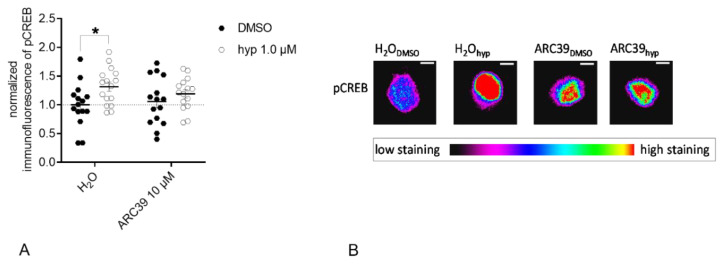
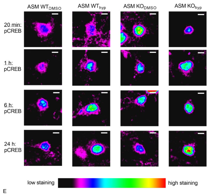
: The acid sphingomyelinase (ASM)/ceramide system exhibits a crucial role in the pathology of major depressive disorder (MDD). ASM hydrolyzes the abundant membrane lipid sphingomyelin to ceramide that regulates the clustering of membrane proteins via microdomain and lipid raft organization. Several commonly used antidepressants, such as fluoxetine, rely on the functional inhibition of ASM in terms of their antidepressive pharmacological effects. Transient receptor potential canonical 6 (TRPC6) ion channels are located in the plasma membrane of neurons and serve as receptors for hyperforin, a phytochemical constituent of the antidepressive herbal remedy St. John's wort. TRPC6 channels are involved in the regulation of neuronal plasticity, which likely contributes to their antidepressant effect. In this work, we investigated the impact of reduced ASM activity on the TRPC6 function in neurons. A lipidomic analysis of cortical brain tissue of ASM deficient mice revealed a decrease in ceramide/sphingomyelin molar ratio and an increase in sphingosine. In neurons with ASM deletion, hyperforin-mediated Ca2+-influx via TRPC6 was decreased. Consequently, downstream activation of nuclear phospho-cAMP response element-binding protein (pCREB) was changed, a transcriptional factor involved in neuronal plasticity. Our study underlines the importance of balanced ASM activity, as well as sphingolipidome composition for optimal TRPC6 function. A better understanding of the interaction of the ASM/ceramide and TRPC6 systems could help to draw conclusions about the pathology of MDD.

Keywords: acid sphingomyelinase; major depressive disorder; sphingolipids; trpc6.


Conflict of interest statement

The authors declare no conflicts of interest.

Figures

Figure 1
Figure 1
Acid sphingomyelinase (ASM) activity is decreased in cortices of ASM KO mice. ASM activity was measured in triplicates using an enzymatic activity assay. Bars indicate mean ± SEM; n = 5–6 animals; **** p ≤ 0.0001.
Figure 2
Figure 2
Genetic ASM deficiency impacts the sphingolipidome in the murine frontal cortex. (A). Concentrations of all analyzed SM species were significantly increased. (B). The concentrations of Cer 16:0, Cer 18:0, Cer 20:0, Cer 22:0, as well as Cer 24:0 were elevated. (C). The overall molar ratio of Cer/SM was decreased in ASM KO mice compared with WT mice. (D). The concentrations of sphingosine were significantly increased in ASM KO mice vs. ASM WT. (E). The concentration of S1P did not show any significant change in ASM KO mice. Bars indicate mean ± SEM; n = 6 animals; * p ≤ 0.05, ** p ≤ 0.01, *** p ≤ 0.001, **** p ≤ 0.0001.
Figure 3
Figure 3
Genetic ASM deficiency does not affect diacylglycerol levels in the murine frontal cortex. (A). Deletion of ASM showed no significant impact on DAG (16:0; 18:1) or (B). DAG (18:0; 20:4) concentrations in frontal cortex. Bars indicate mean ± SEM; n = 6 animals.
Figure 4
Figure 4
Genetic ASM deficiency decreases TRPC6-mediated Ca2+ influx in synaptosomes and cultured primary murine neuronal cells. (A). Hyperforin-induced (10 µM) Ca2+ influx in murine brain synaptosomes, isolated from ASM KO mice, was reduced compared with synaptosomes isolated from ASM WT mice (n = 6 animals). (B). Application of hyperforin 10 µM activates TRPC6-mediated Ca2+ influx. The amplitude of change in intracellular Ca2+ concentration is diminished in cortical ASM KO neurons compared with cortical ASM WT. (C). Representative time-curve of TRPC6-mediated Ca2+ influx in cortical mouse neurons. Arrow indicates the application of hyperforin 10 µM. (D). Baseline Ca2+ concentrations of ASM KO and ASM WT cortical neurons do not differ. Bars indicate mean ± SEM; n = 4 cell batches, 1–4 cover slips per batch were analyzed; ** p ≤ 0.01, *** p ≤ 0.001.
Figure 5
Figure 5
Co-culturing of ASM WT glial cells with ASM KO neurons does not rescue TRPC6 phenotype. (A). Ca2+ influx is reduced in ASM-deficient cortical mouse neurons, which were grown together with ASM WT glial cells compared with ASM WT neurons, which were grown with ASM WT glial cells in a Banker’s culture. (B). Representative time-curve of TRPC6-mediated Ca2+ influx in cortical mouse neurons, which were grown in a Banker’s culture with ASM WT glial cells. Arrow indicates the application of hyperforin 10 µM. Bars indicate mean ± SEM; n = 3 cell batches, 1–2 cover slips per batch were analyzed; * p ≤ 0.05.
Figure 6
Figure 6
ASM activity inhibition prevents hyperforin-induced CREB phosphorylation in primary rat neuronal cells. (A). Hyperforin (1 µM) induced CREB phosphorylation in rat excitatory cortical neurons after 24 h of treatment. This effect was partly abolished when ASM activity was inhibited with ARC39 10 µM (B). Representative images of pCREB staining in single nuclei of excitatory rat neurons after indicated treatments. Scale bar indicates 5 µm. Bars indicate mean ± SEM; n = 1 cell batch, 3 cover slips analyzed per batch, 5 regions analyzed per cover slip; * p ≤ 0.05.
Figure 7
Figure 7
Genetic ASM deficiency impacts hyperforin-induced CREB-phosphorylation in excitatory primary murine neuronal cells. (A). 24 h after hyperforin (1 µM) stimulation, both ASM WT and ASM KO excitatory neurons showed a significant increase in pCREB staining compared with control conditions. (B). After a 20 min treatment of ASM KO and ASM WT neurons with hyperforin, distinctive hyperforin-induced changes were visible. While in ASM WT neurons after hyperforin application, a small positive trend was visible towards stronger pCREB staining, ASM KO neurons contrarily showed significantly decreased pCREB levels. (C). 1 h after hyperforin application, no treatment or genotype-induced effects were observed. (D). 6 h after hyperforin stimulation, no treatment or genotype-induced effects were observed. (E) Representative images of pCREB staining in single nuclei of excitatory murine neurons after indicated treatments and time points. Scale bar indicates 5 µm. Bars indicate mean ± SEM; n = 2 cell batches, 1 cover slip analyzed per batch, 3–7 regions analyzed per cover slip; * p ≤ 0.05, ** p ≤ 0.01.
Figure 8
Figure 8
Genetic ASM deficiency impacts hyperforin-induced CREB-phosphorylation in inhibitory primary murine neuronal cells. (A). 24 h after hyperforin (1 µM) stimulation, ASM KO inhibitory neurons showed a significant increase in pCREB staining, ASM WT neurons a trend towards an increase. (B). After a 20 min treatment, ASM WT neurons showed a small positive trend towards stronger pCREB staining, and ASM KO neurons contrarily showed significantly decreased pCREB levels. (C). 1 h after hyperforin application, no treatment or genotype-induced effects were observed. (D). 6 h after hyperforin stimulation, no treatment or genotype-induced effects were observed. (E). Representative images of pCREB staining in single nuclei of excitatory murine neurons after indicated treatments and time points. Scale bar indicates 5 µm. Bars indicate mean ± SEM; n = 2 cell batches, 1 cover slip analyzed per batch, 3–7 regions analyzed per cover slip; * p ≤ 0.05, *** p ≤ 0.001.
Figure 8
Figure 8
Genetic ASM deficiency impacts hyperforin-induced CREB-phosphorylation in inhibitory primary murine neuronal cells. (A). 24 h after hyperforin (1 µM) stimulation, ASM KO inhibitory neurons showed a significant increase in pCREB staining, ASM WT neurons a trend towards an increase. (B). After a 20 min treatment, ASM WT neurons showed a small positive trend towards stronger pCREB staining, and ASM KO neurons contrarily showed significantly decreased pCREB levels. (C). 1 h after hyperforin application, no treatment or genotype-induced effects were observed. (D). 6 h after hyperforin stimulation, no treatment or genotype-induced effects were observed. (E). Representative images of pCREB staining in single nuclei of excitatory murine neurons after indicated treatments and time points. Scale bar indicates 5 µm. Bars indicate mean ± SEM; n = 2 cell batches, 1 cover slip analyzed per batch, 3–7 regions analyzed per cover slip; * p ≤ 0.05, *** p ≤ 0.001.

References

    1. Gulbins E., Palmada M., Reichel M., Lüth A., Böhmer C., Amato D., Müller C.P., Tischbirek C.H., Groemer T.W., Tabatabai G., et al. Acid sphingomyelinase–ceramide system mediates effects of antidepressant drugs. Nat. Med. 2013;19:934–938. doi: 10.1038/nm.3214. - DOI - PubMed
    1. Kornhuber J., Medlin A., Bleich S., Jendrossek V., Henkel A.W., Wiltfang J., Gulbins E. High activity of acid sphingomyelinase in major depression. J. Neural Transm. 2005;112:1583–1590. doi: 10.1007/s00702-005-0374-5. - DOI - PubMed
    1. Kornhuber J., Müller C.P., Becker K.A., Reichel M., Gulbins E. The ceramide system as a novel antidepressant target. Trends Pharmacol. Sci. 2014;35:293–304. doi: 10.1016/j.tips.2014.04.003. - DOI - PubMed
    1. Schneider P.B., Kennedy E.P. Sphingomyelinase in normal human spleens and in spleens from subjects with Niemann-Pick disease. J. Lipid Res. 1967;8:202–209. - PubMed
    1. Grassmé H., Riethmüller J., Gulbins E. Biological aspects of ceramide-enriched membrane domains. Prog. Lipid Res. 2007;46:161–170. doi: 10.1016/j.plipres.2007.03.002. - DOI - PubMed

Publication types

Substances

LinkOut - more resources