Review
. 2021 Jan;16(1):99-117.
doi: 10.1016/j.cpet.2020.09.012.

Total-Body PET Imaging of Musculoskeletal Disorders

Affiliations
Review

Total-Body PET Imaging of Musculoskeletal Disorders

Abhijit J Chaudhari et al. PET Clin. 2021 Jan.

Abstract

Imaging of musculoskeletal disorders, including arthritis, infection, osteoporosis, sarcopenia, and malignancies, is often limited when using conventional modalities such as radiography, computed tomography (CT), and MR imaging. As a result of recent advances in Positron Emission Tomography (PET) instrumentation, total-body PET/CT offers a longer axial field-of-view, higher geometric sensitivity, and higher spatial resolution compared with standard PET systems. This article discusses the potential applications of total-body PET/CT imaging in the assessment of musculoskeletal disorders.

Keywords: Arthritis; Cancer; Fever of unknown origin; Osteoporosis; Osteosarcoma; PET/CT; PET/MR imaging; Sarcopenia.


Conflict of interest statement

Disclosure UC Davis has a revenue-sharing agreement with United Imaging Healthcare. The authors have no other matters to disclose. Conflict of interest The authors have declared no conflicts of interest.

Figures

Figure 1
Figure 1
TB PET in rheumatoid and psoriatic arthritis. Maximum intensity projection (MIP) from the TB PET scan of a 65-year-old man with established rheumatoid arthritis (A). Images of the subject in (A) showing classic ring-like patterns of radiotracer uptake consistent with synovitis in the joints of the hand (B), and foot (C). Extensor and flexor tenosynovitis and enthesitis in the right second digit of a 72-year-old man with established psoriatic arthritis (D), also shown in the MIP of the TB PET scan (E). These images were acquired on the uEXPLORER PET/CT scanner at the University of California Davis, with an injected dose of about 74 MBq. Images show static scans conducted over 20 min, starting at 40 min post-radiotracer injection. Courtesy of Y. Abdelhafez, MD, University of California Davis
Figure 2
Figure 2
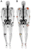
FDG-PET maximum intensity projection (MIP) of a 69-year-old man with rheumatoid arthritis showing the upper body (A) and lower body (B). Synovitis was assessed by segmenting FDG-avid joints using an adaptive thresholding algorithm (ROVER software, ABX GmbH, Radeberg, Germany). Metabolically active volume (MAV), maximum standardized uptake value (SUVmax), mean SUV (SUVmean), partial volume-corrected SUVmean (pvcSUVmean), total lesion glycolysis (TLG = MAV × SUVmean), and partial volume-corrected TLG (pvcTLG = MAV × pvcSUVmean) were calculated and summed for each segmented region. The global pvcTLG for this patient was 820.0. These analyses could be enabled at a lower radiation dose and scan time, with more comprehensive body coverage in a single scan, using TB PET/CT. From Saboury B, Morris MA, Nikpanah M, Werner TJ, Jones EC, Alavi A. Reinventing Molecular Imaging with Total-Body PET, Part II: Clinical Applications. PET Clin. 2020;15(4):463-475. doi:10.1016/j.cpet.2020.06.013; with permission.
Figure 3
Figure 3
NaF-PET maximum intensity projection (MIP) of the same rheumatoid arthritis patient as Figure 2 showing the upper body (A) and lower body (B). ROVER software was used to segment focal areas of high bone formation in the joints. Metabolically active volume (MAV), maximum standardized uptake value (SUVmax), mean SUV (SUVmean), partial volume-corrected SUVmean (pvcSUVmean), total calcium metabolism (TCM = MAV × SUVmean), and partial volume-corrected TCM (pvcTCM = MAV × pvcSUVmean) were calculated and summed for each segmented region. The global pvcTCM for this patient was 1898.8. From Saboury B, Morris MA, Nikpanah M, Werner TJ, Jones EC, Alavi A. Reinventing Molecular Imaging with Total-Body PET, Part II: Clinical Applications. PET Clin. 2020;15(4):463-475. doi:10.1016/j.cpet.2020.06.013
Figure 4
Figure 4
NaF-PET maximum intensity projection (MIP) of a 72-year-old man showing tracer uptake in the joints before (A) and after (B) PET segmentation using an adaptive thresholding algorithm (ROVER software). Volumetric and metabolic parameters were automatically calculated and summed to determine total disease activity. The global partial volume-corrected total calcium metabolism for this patient was 338.6.
Figure 5
Figure 5
Whole-body FDG-PET/CT images of a 60-year-old man with multiple myeloma. The cortical bone and bone marrow were segmented using a growing region algorithm based on Hounsfield units, followed by smoothing and closing algorithms (OsiriX software; Pixmeo SARL; Bernex, Switzerland). The global SUVmean, which represents the whole bone marrow activity, before initiating treatment (A) was 2.02 and decreased to 1.10 after finishing the course of treatment (B). (From Raynor WY, Al-Zaghal A, Zadeh MZ, Seraj SM, Alavi A. Metastatic Seeding Attacks Bone Marrow, Not Bone: Rectifying Ongoing Misconceptions. PET Clin. 2019;14(1): 135-144; with permission)
Figure 6
Figure 6
Maximum intensity projection (MIP) of the CT image (A) showing a methodology used to segment muscle. Two lines (horizontal parallel green lines) corresponding to 5 cm above the intercondylar notch and 5 cm below the greater trochanter were manually delineated according to pre-determined anatomical criteria. A growing region algorithm with lower and upper thresholds of 1 and 150 Hounsfield units, respectively, was used to segment the muscle (OsiriX software) (B). Applying this methodology to 71 subjects, thigh muscle volume was found to decrease with age, and uptake of FDG uptake was found to be significantly higher on the right side compared to the left. (From Kothekar E, Yellanki D, Borja AJ, et al. 18F-FDG-PET/CT in measuring volume and global metabolic activity of thigh muscles: a novel CT-based tissue segmentation methodology. Nucl Med Commun. 2020;41(2):162-168.; with permission)
Figure 7
Figure 7
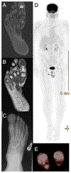
A 58-year-old man with toe pain was evaluated with MRI of the foot. A marrow-replacing lesion of the first distal phalanx with T2 hyper- (A) and T1 hypo-signal intensity (B) was identified, the tissue sampling of which was consistent with osteosarcoma. After surgical resection and several courses of chemotherapy (C), the patient returned for restaging with whole-body FDG-PET/CT. A hypermetabolic right ankle (Kager fat pad, D, E) and inguinal lymph nodes (D) with respective SUVmax of 4.4 and 10.4 were identified, in keeping with metastatic lymphadenopathy.

References

    1. Briggs AM, Cross MJ, Hoy DG, et al. Musculoskeletal Health Conditions Represent a Global Threat to Healthy Aging: A Report for the 2015 World Health Organization World Report on Ageing and Health. Gerontologist. 2016;56 Suppl 2:S243–255. - PubMed
    1. Murray CJ, Barber RM, Foreman KJ, et al. Global, regional, and national disability-adjusted life years (DALYs) for 306 diseases and injuries and healthy life expectancy (HALE) for 188 countries, 1990–2013: quantifying the epidemiological transition. The Lancet. 2015;386(10009):2145–2191. - PMC - PubMed
    1. March L, Smith EU, Hoy DG, et al. Burden of disability due to musculoskeletal (MSK) disorders. Best Practice & Research: Clinical Rheumatology. 2014;28(3):353–366. - PubMed
    1. Put S, Westhovens R, Lahoutte T, Matthys P. Molecular imaging of rheumatoid arthritis: emerging markers, tools, and techniques. Arthritis research & therapy. 2014;16(2):208. - PMC - PubMed
    1. Gholamrezanezhad A, Guermazi A, Salavati A, Alavi A. Evolving Role of PET-Computed Tomography and PET-MR Imaging in Assessment of Musculoskeletal Disorders and Its Potential Revolutionary Impact on Day-to-Day Practice of Related Disciplines. PET Clin. 2018;13(4):xiii–xiv. - PubMed

MeSH terms