

Three Cases of COVID-19 Pneumonia in Female Patients in Italy Who Had Pulmonary Fibrosis on Follow-Up Lung Computed Tomography Imaging
- PMID: 33219200
- PMCID: PMC7690330
- DOI: 10.12659/AJCR.926921
Three Cases of COVID-19 Pneumonia in Female Patients in Italy Who Had Pulmonary Fibrosis on Follow-Up Lung Computed Tomography Imaging
Abstract
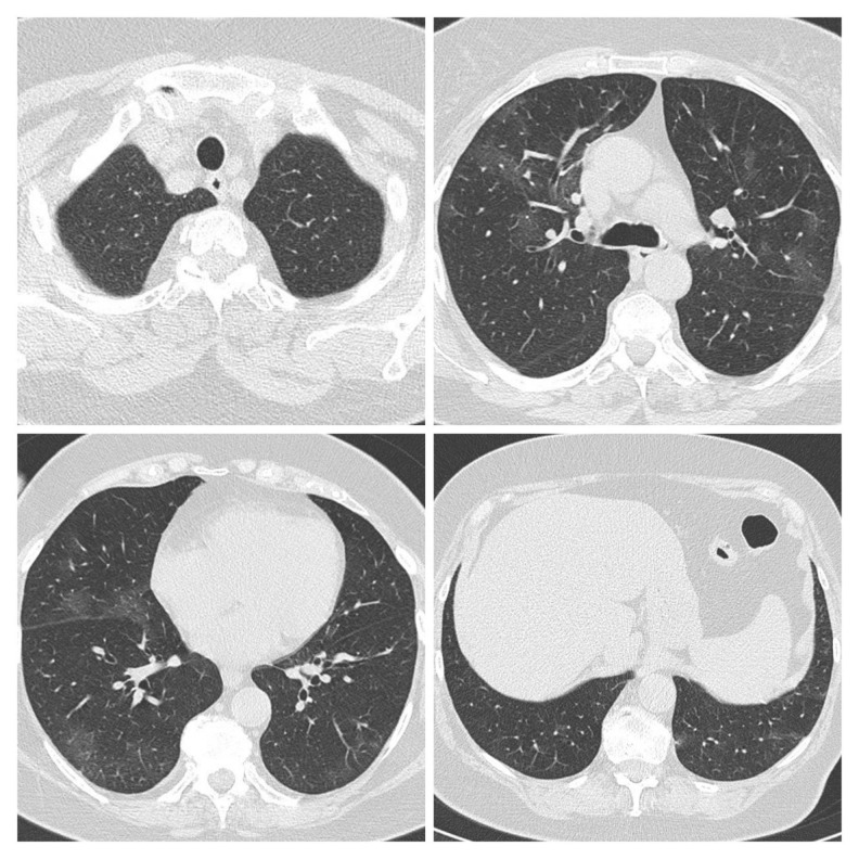
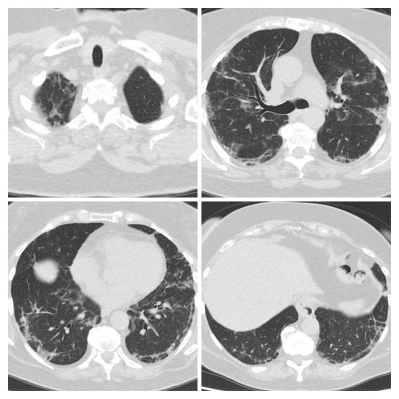
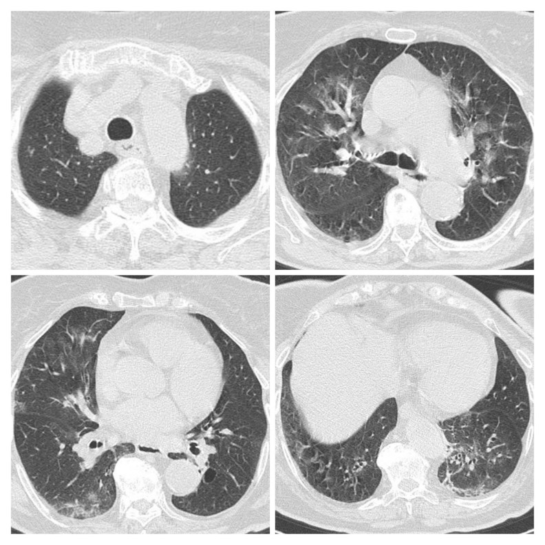
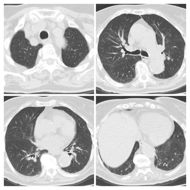
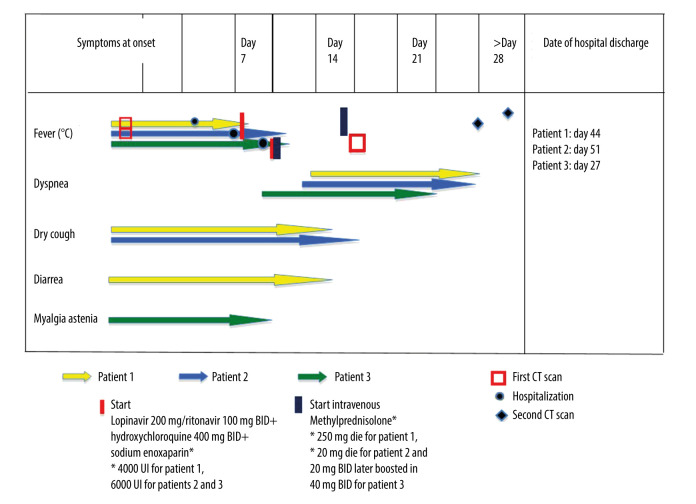
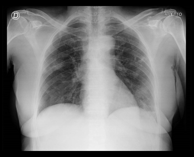
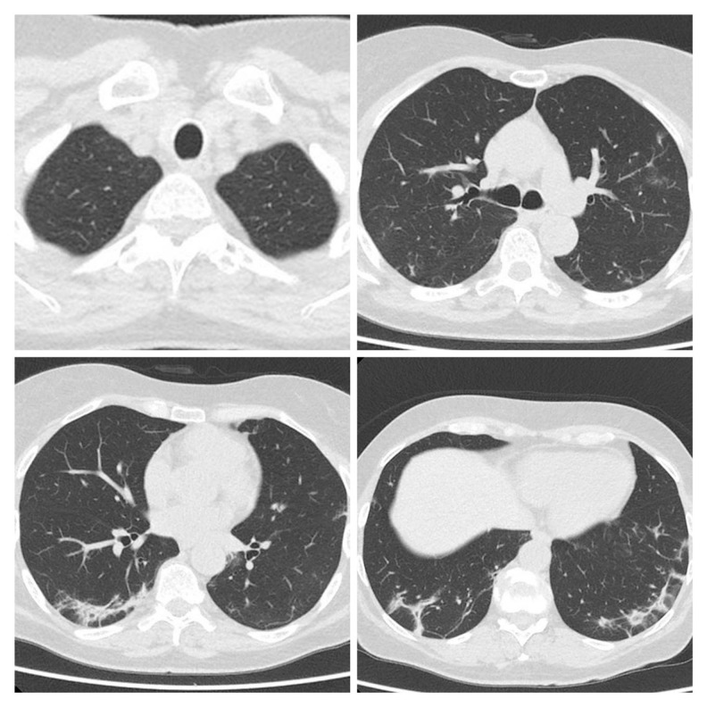
BACKGROUND Since December 2019, an outbreak caused by a novel coronavirus infection (severe acute respiratory syndrome coronavirus 2, SARS-CoV-2) occurred in Wuhan, China, and it rapidly spread all over the world. The clinical spectrum of coronavirus disease 2019 (COVID-19) is wide, with acute respiratory distress syndrome (ARDS) occurring in 15% of patients affected, requiring high oxygen support. Currently, there is no clearly effective antiviral therapy. Steroids and immunomodulators are under investigation for potential activity. Little is known about middle and long-term sequelae on respiratory function. According to some authors, COVID-19 could cause pulmonary fibrosis. We report 3 cases of pulmonary fibrosis detected on follow-up computed tomography (CT) imaging in 3 female patients who recovered from COVID-19 pneumonia in Italy (L'Aquila, Abruzzo). CASE REPORT All patients were female and had no significant previous respiratory disease or history of smoke exposure, and none had received high-flow oxygen support during treatment of the disease. In all cases, late onset of mild dyspnea, slow and incomplete respiratory recovery, and early evidence of fibrous signs on chest CT scan were characteristic of the clinical course. CONCLUSIONS This report focuses on a possible scenario of long-term lung damage in COVID-19 pneumonia survivors. Limitations are lack of long-term follow-up and functional data in the very early phase. It is advantageous that all COVID-19 pneumonia patients undergo serial chest CT and spirometry long-term follow-up for at least 1 year to assess residual damage. This is particularly relevant in those with slow respiratory recovery and long hospitalization.
Conflict of interest statement
Figures
References
-
- World Health Organization (WHO) Coronavirus disease 2019 (COVID-19) Situation Report-92. 2020. https://www.who.int/docs/default-source/coronaviruse/situation-reports/2....
-
- Ward PA, Hunninghake GW. Lung inflammation and fibrosis. Am J Respir Crit Care Med. 1998;157(4 Pt 2):S123–29. - PubMed
Publication types
MeSH terms
LinkOut - more resources
Full Text Sources
Medical
Miscellaneous
