. 2020 Nov 20;10(1):20258.
doi: 10.1038/s41598-020-76927-0.

CMT-3 targets different α-synuclein aggregates mitigating their toxic and inflammogenic effects

Affiliations

CMT-3 targets different α-synuclein aggregates mitigating their toxic and inflammogenic effects

Florencia González-Lizárraga et al. Sci Rep. .

Abstract

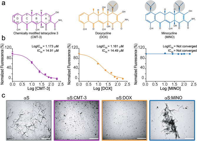
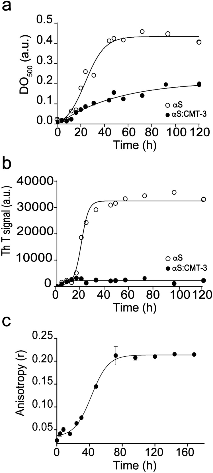
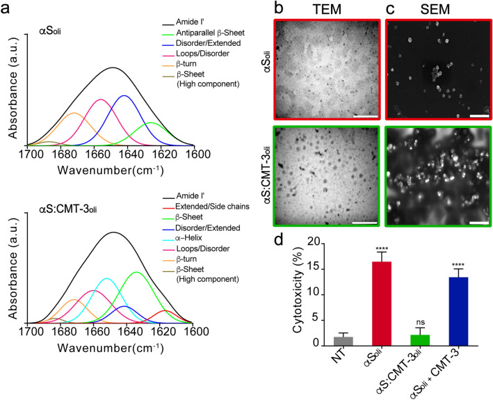
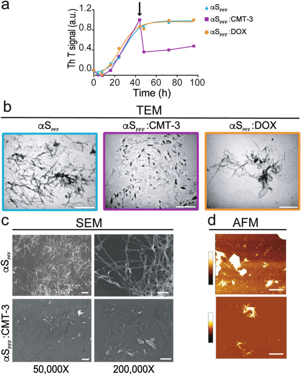
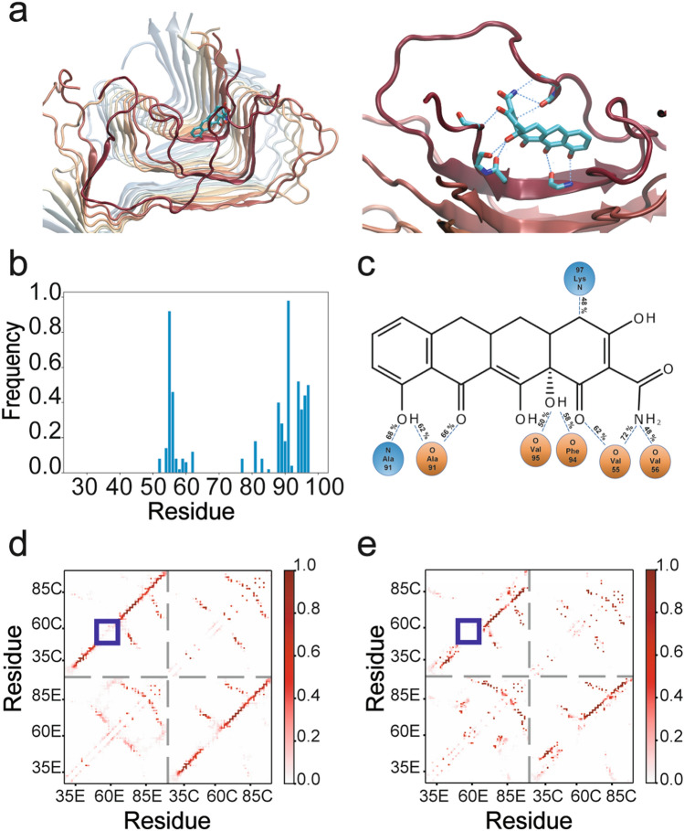
Parkinson's disease (PD) is a neurodegenerative disorder for which only symptomatic treatments are available. Repurposing drugs that target α-synuclein aggregation, considered one of the main drivers of PD progression, could accelerate the development of disease-modifying therapies. In this work, we focused on chemically modified tetracycline 3 (CMT-3), a derivative with reduced antibiotic activity that crosses the blood-brain barrier and is pharmacologically safe. We found that CMT-3 inhibited α-synuclein amyloid aggregation and led to the formation of non-toxic molecular species, unlike minocycline. Furthermore, CMT-3 disassembled preformed α-synuclein amyloid fibrils into smaller fragments that were unable to seed in subsequent aggregation reactions. Most interestingly, disaggregated species were non-toxic and less inflammogenic on brain microglial cells. Finally, we modelled the interactions between CMT-3 and α-synuclein aggregates by molecular simulations. In this way, we propose a mechanism for fibril disassembly. Our results place CMT-3 as a potential disease modifier for PD and possibly other synucleinopathies.


Conflict of interest statement

The authors declare no competing interests.

Figures

Figure 1
Figure 1
Comparative effect of CMT-3, DOX and MINO on α-synuclein amyloid aggregation. (a) Chemical structures of CMT-3, DOX, and MINO. Atom numbering and ring labels of the tetracycline skeleton are provided on the chemical structure of CMT-3. Note that the main difference in these structures resides in the presence or not of DMA groups designated by grey shaded circles. (b) Dose–response curves from endpoint ThT intensities of α-synuclein aggregation assay in the presence of different concentrations of CMT-3, DOX or MINO. IC50 values are mean values of three independent determinations. The data fitting is described in “Methods” section. (c) TEM of α-synuclein samples incubated at 37 °C for 120 h under continuous orbital agitation in the absence (αS) or in presence of CMT-3 (αS:CMT-3), DOX (αS:DOX), or MINO (αS:MINO). The scale bar corresponds to 1 μm, at ×26,000 magnification.
Figure 2
Figure 2
CMT-3 inhibits α-synuclein amyloid aggregation by binding to aggregated species. (a) Turbidimetry assay of 70-μM α-synuclein samples incubated in the absence (empty circles) or presence of 100 μM CMT-3 (full circles) and monitored at 500 nm. (b) ThT fluorescence of 70 μM α-synuclein samples incubated in the absence (empty circles) or presence of 100 μM CMT-3 (full circles). (c) Binding of CMT-3 to α-synuclein aggregated species monitored by fluorescence anisotropy. Changes in fluorescence anisotropy of CMT-3 were measured at λem 520 nm upon the addition of aliquot samples harvested at different time-points of an α-synuclein aggregation reaction. Data values represent the mean ± S.E. of 6 independent experiments.
Figure 3
Figure 3
CMT-3 reshapes α-synuclein early aggregates into non-toxic species. (a) Analysis of α-synuclein Amide I' band after the curve fitting procedure (see “Methods”) showing the component bands: αS in the absence (top) or in the presence of CMT-3 (bottom) after 16 h incubation. (b) TEM and (c) SEM of α-synuclein samples incubated at 37 °C under orbital agitation in the absence (top) or in the presence of CMT-3 (bottom), and harvested after 16 h for observation of oligomers. The white bar corresponds to 500 nm. (d) LDH cytotoxicity assay in SH-SY5Y cells after the addition of α-synuclein oligomers formed after 16 h of incubation at 37 °C under orbital agitation in the absence (αSoli) or presence of CMT-3 (αS:CMT-3oli). Cytotoxicity values were normalized by the signal observed after addition of Triton X-100, which induced complete disruption of the cells. Data represents the mean ± S.E.M (n = 11). One-way ANOVA followed by Holm-Sidak’s multiple comparisons test. ****p < 0.0001 vs NT.
Figure 4
Figure 4
CMT-3, unlike DOX, disassembles preformed α-synuclein amyloid fibrils (αSPFF). (a) In ThT fluorescence assays, 70 μM α-synuclein was either incubated alone (blue line), or with 100 μM of CMT-3 (purple line) or DOX (orange line) added 44 h after the initiation of the aggregation process (arrow). (b) TEM of α-synuclein samples incubated at 37 °C under orbital agitation during 44 h to obtain fibrillar species (αSPFF). Samples were further incubated for another 52 h either alone or with the addition of 100 μM CMT-3 or DOX. Note the dramatic reduction in αSPFF with the addition of CMT-3, but not with DOX. The scale bar corresponds to 1 μm (×26,000). (c, d) αSPFF and αSPFF:CMT-3 species from (b) were further submitted to SEM and AFM. SEM scale bar corresponds to 500 nm (×50,000) and 200 nm (×200,000). AFM scale bar corresponds to 1 μm. The pseudo color scale bar represents the information of the z axis from 0 to 100 nm. The z range was chosen to allow the observation of fibrils while the aggregated fibrillar clusters are out of range since they have a height greater than 100 nm.
Figure 5
Figure 5
Disassembled α-synuclein PFF (αSPFF:CMT-3) are non-toxic and unable to either disrupt membrane integrity or promote seeding. (a) In seeding assays, αSPFF served as efficient seeds to accelerate aggregation of α-synuclein monomers (light blue squares), while αSPFF:CMT-3 were not only unable to seed monomers (purple triangles), but also blocked the amyloid aggregation reaction of the monomers. The unseeded aggregation kinetics of αSm (no seeds) is also shown (black circles). For this assay, solutions were incubated at 37 °C under continuous orbital agitation and aggregation was measured by ThT fluorescence emission. (b) Changes in liposomal membrane permeability upon the addition of α-synuclein monomers (αSm), α-synuclein oligomers harvested after 16 h of incubation (αSoli), αSPFF or αSPFF:CMT-3. Results show that new species produced by CMT-3-driven disassembly of αSPFF were unable to disrupt membrane permeability. The fluorescence signal was normalized to values obtained after addition of Triton X-100, which induced complete disruption of liposomal vesicles. (c) LDH cytotoxicity assay in SH-SY5Y cells after the addition of αSm, αSoli, αSPFF and αSPFF:CMT-3. The cytotoxicity signal was normalized to values obtained after addition of Triton X-100. Data represents the mean ± S.E.M (n = 11). One-way ANOVA followed by Holm-Sidak’s multiple comparisons test. ****p < 0.0001 vs NT.
Figure 6
Figure 6
Disassembled α-synuclein PFF (αSPFF:CMT-3) are less inflammogenic for microglial cells. (a) TNF-α release in microglial cells treated for 24 h with αSPFF, αSPFF:CMT-3 (70 μg/ml), or intact αSPFF in wells pre-incubated with CMT-3 (CMT-3preinc + αSPFF). (b) Quantification of glutamate release after exposure of microglial cultures to the same treatments as in (a). Data represents the mean ± S.E.M (n = 11). One-way ANOVA followed by Holm-Sidak’s multiple comparisons test. *p < 0.05, **p < 0.01, ****p < 0.0001 vs NT. #p < 0.05, #### p < 0.0001 vs αSPFF..
Figure 7
Figure 7
Binding mode of CMT-3 to α-synuclein fibrils and subsequent disassembly mechanism (a) Side and close-up views of the putative complex formed between CMT-3 and α-synuclein fibrils. The protein backbone is represented in cartoon while the ligand is depicted in ball-and-stick representation. The hydrogen bond network between the protein and the ligand is represented as a discontinued blue line in the enlarged side view. (b) Contact frequency between CMT-3 and α-synuclein amino acid residues. (c) Schematic representation of the hydrogen bonds between CMT-3 and atoms from amide and carbonyl groups in the main chain of α-synuclein. Contact map between the residues at the edge (E) or the core (C) of the α-synuclein fiber in the absence (d) or presence of CMT-3 (e). The color bar scale represents the frequency contact along the simulation time.

References

    1. GBD 2016 Parkinson’s Disease Collaborators Global, regional, and national burden of Parkinson’s disease, 1990–2016: A systematic analysis for the Global Burden of Disease Study 2016. Lancet. Neurol. 2018;17:939–953. doi: 10.1016/S1474-4422(18)30295-3. - DOI - PMC - PubMed
    1. Fereshtehnejad S-M, Zeighami Y, Dagher A, Postuma RB. Clinical criteria for subtyping Parkinson’s disease: Biomarkers and longitudinal progression. Brain. 2017;140:1959–1976. doi: 10.1093/brain/awx118. - DOI - PubMed
    1. Picconi B, Hernández LF, Obeso JA, Calabresi P. Motor complications in Parkinson’s disease: Striatal molecular and electrophysiological mechanisms of dyskinesias. Mov. Disord. 2018;33:867–876. doi: 10.1002/mds.27261. - DOI - PubMed
    1. Spillantini MG, et al. Alpha-synuclein in Lewy bodies. Nature. 1997;388:839–840. doi: 10.1038/42166. - DOI - PubMed
    1. Araki K, et al. Parkinson’s disease is a type of amyloidosis featuring accumulation of amyloid fibrils of α-synuclein. Proc. Natl. Acad. Sci. U. S. A. 2019;116:17963–17969. doi: 10.1073/pnas.1906124116. - DOI - PMC - PubMed

Publication types