Review
. 2021 Jan;17(1):37-51.
doi: 10.1038/s41582-020-00427-y. Epub 2020 Nov 20.

Progressive multifocal leukoencephalopathy and the spectrum of JC virus-related disease

Affiliations
Review

Progressive multifocal leukoencephalopathy and the spectrum of JC virus-related disease

Irene Cortese et al. Nat Rev Neurol. 2021 Jan.

Abstract

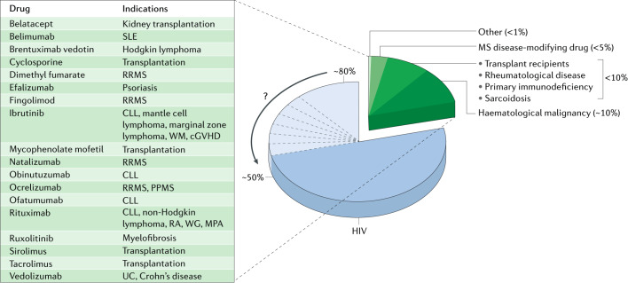
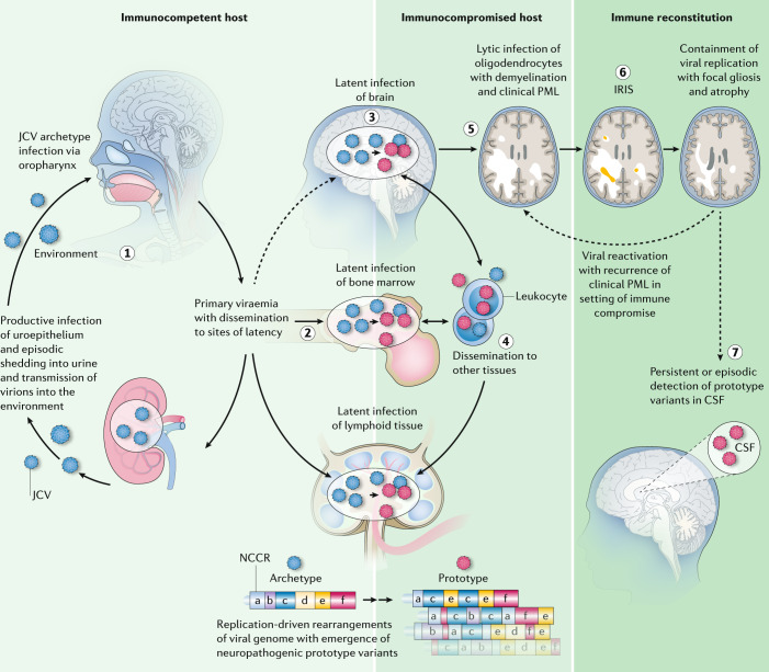
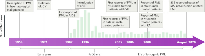
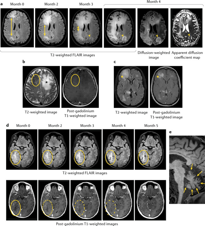
Progressive multifocal leukoencephalopathy (PML) is a devastating CNS infection caused by JC virus (JCV), a polyomavirus that commonly establishes persistent, asymptomatic infection in the general population. Emerging evidence that PML can be ameliorated with novel immunotherapeutic approaches calls for reassessment of PML pathophysiology and clinical course. PML results from JCV reactivation in the setting of impaired cellular immunity, and no antiviral therapies are available, so survival depends on reversal of the underlying immunosuppression. Antiretroviral therapies greatly reduce the risk of HIV-related PML, but many modern treatments for cancers, organ transplantation and chronic inflammatory disease cause immunosuppression that can be difficult to reverse. These treatments - most notably natalizumab for multiple sclerosis - have led to a surge of iatrogenic PML. The spectrum of presentations of JCV-related disease has evolved over time and may challenge current diagnostic criteria. Immunotherapeutic interventions, such as use of checkpoint inhibitors and adoptive T cell transfer, have shown promise but caution is needed in the management of immune reconstitution inflammatory syndrome, an exuberant immune response that can contribute to morbidity and death. Many people who survive PML are left with neurological sequelae and some with persistent, low-level viral replication in the CNS. As the number of people who survive PML increases, this lack of viral clearance could create challenges in the subsequent management of some underlying diseases.


Conflict of interest statement

The authors declare no competing interests.

Figures

Fig. 1
Fig. 1. Risk factors for PML.
The chart shows the estimated proportion of progressive multifocal leukoencephalopathy (PML) cases that occur in different patient groups. PML is not a reportable disease, so accurate estimates of incidence among patient populations are lacking. Over the past four decades, HIV infection has accounted for the vast majority of PML cases (~80%), although evidence suggests that the proportion is shifting towards lower numbers, as indicated by the arrow. Approximately 10% of PML cases occur among patients with haematological malignancy, most commonly non-Hodgkin lymphoma and chronic lymphocytic leukaemia (CLL). Natalizumab-associated PML accounts for the third-largest single patient population, estimated at less than 5% of cases. Approximately 10% of PML cases occur in other patients with identifiable immunosuppressed states, including transplant recipients and patients with underlying chronic inflammatory or rheumatological disease. In rare cases, patients without a clearly identifiable predisposing condition develop PML; this occurs more commonly among elderly patients. The inset table shows the growing number of drugs associated with a risk of PML and that currently carry a labelled warning; such medications often contribute to the risk of PML among susceptible patient populations. cGVHD, chronic graft-versus-host disease; MPA, microscopic polyangiitis; MS, multiple sclerosis; PPMS, primary progressive multiple sclerosis; RA, rheumatoid arthritis; RRMS, relapsing–remitting multiple sclerosis; SLE, systemic lupus erythematosus; UC, ulcerative colitis; WG, Wegener granulomatosis; WM, Waldenstrom macroglobulinaemia.
Fig. 2
Fig. 2. Pathophysiology of PML.
According to the three-stage model of progressive multifocal leukoencephalopathy (PML) described by Pavlovic et al., the first stage of infection, which occurs in most of the general population, is subclinical, persistent infection of the kidney with non-pathogenic archetype JC virus (JCV). Shedding of virus into the urine is probably the major source of environmental contamination (step 1). During this stage, the virus can spread to secondary sites, including the bone marrow, lymphoid tissue and possibly the brain, where latent infection can be established (step 2). The second stage of infection can occur with prolonged impairment of cellular immunity, which allows viral reactivation and accumulation of neuropathogenic JCV variants with rearrangements of the non-coding control region (NCCR) (step 3). The anatomic sites of these mutational events are not known. Leukocytes, including CD34+ progenitor cells, B cell lineage cells and other lymphocyte subsets could be vehicles for haematogenous spread of JCV, either via infection of these cells or via passive transportation of virus on the cell surface (step 4). Neurotropic virus might reach the brain at this stage via transportation by leukocytes or as cell-free virus, although local transformation from latent archetype virus has not been excluded. The third stage is clinically manifest PML (step 5). On immune reconstitution, as a result of either specific treatment for PML or discontinuation of an immunosuppressive therapy, subclinical or clinical immune reconstitution inflammatory syndrome (IRIS) can occur (step 6). Patient survival after successful immune reconstitution suggests a fourth stage in which host–virus equilibrium is re-established and latent or persistent infection with rearranged pathogenic virus is established in the brain (step 7). In some patients, low copy numbers of the virus persist in the cerebrospinal fluid (CSF). The risk of PML recurrence in these patients is unknown but it is not negligible. Figure adapted with permission from ref., Elsevier.
Fig. 3
Fig. 3. Timeline of PML disease eras.
From an epidemiological perspective, three distinct eras of progressive multifocal leukoencephalopathy (PML) have been identified. In the early years, following its first description, PML was rare and almost uniformly fatal, seen most commonly in patients with haematological malignancy. During the AIDS pandemic, the incidence of PML increased dramatically — up to 5% of patients with AIDS developed the disease. With the introduction of combined antiretroviral therapies (cART) in 1996, this proportion declined substantially, and immune reconstitution was established as a way to prevent PML and to increase the chance of survival. Although the majority of PML cases worldwide are still associated with AIDS, iatrogenic PML is linked to a growing number of therapies that target immune function. Most notable of these are natalizumab for the treatment of relapsing–remitting multiple sclerosis (MS), rituximab for the treatment of haematological cancers and rheumatological disorders, and efalizumab for the treatment of psoriasis (efalizumab was withdrawn from the market in 2009 after reports of association with PML). JCV, JC virus; RA, rheumatoid arthritis; SLE, systemic lupus erythematosus.
Fig. 4
Fig. 4. MRI findings in various presentations of JC virus infection.
a | MRI of a 40-year-old woman with a history of follicular lymphoma who was treated with bendamustine and rituximab and developed progressive multifocal leukoencephalopathy (PML) while receiving rituximab maintenance therapy. MRI shows progression of PML lesions over a 4-month period. T2-weighted fluid-attenuated inversion recovery (FLAIR) images show enlargement of lesions in the right frontal and parietal lobes (double-headed arrows). At the final time point (month 4), involvement of the right cerebral hemisphere is nearly complete and involvement of the left frontal and parietal lobes is substantial. Mild diffusion restriction (arrow heads) can be seen as hyperintensity on the diffusion-weighted image and as relative hypointensity on the apparent diffusion coefficient map, at the edge of the lesion near the right lateral ventricle and, to a lesser extent, in the left parietal lobe. b | MRI of a 42-year-old man with HIV-related PML after initiation of treatment with antiretroviral therapy. The ‘Milky Way’ appearance, an MRI feature first described in natalizumab-related PML but that can generally be observed in inflammatory PML, is apparent in these T2-weighted and post-gadolinium T1-weighted images, which show punctate foci of signal abnormality (circled) in addition to the large, non-enhanced lesion in the left frontal lobe (asterisk). c | MRI of a 61-year-old man with HIV-related PML who developed immune reconstitution inflammatory syndrome (IRIS) after initiation of treatment with antiretroviral therapy. MRI shows oedematous lesions in the right frontal lobe and corpus callosum (asterisks). d | MRI of a 34-year-old man with idiopathic lymphopenia. MRI shows shrinkage of a right temporal lobe lesion over 5 months after successful immune reconstitution with checkpoint inhibition. T2-weighted FLAIR images show reduction in the lesion size (top row, circled) and post-gadolinium T1-weighed images show gradual resolution of contrast enhancement (dashed rings), although resolution of contrast enhancement does not indicate resolution of the lesion. e | MRI of a 60-year-old man with HIV and JC virus granule cell neuronopathy. T1-weighted MRI shows hallmark profound cerebellar atrophy (arrows).

Similar articles

Cited by

References

    1. Padgett BL, Walker DL, ZuRhein GM, Eckroade RJ, Dessel BH. Cultivation of papova-like virus from human brain with progressive multifocal leucoencephalopathy. Lancet. 1971;1:1257–1260. - PubMed
    1. Moens U, et al. Biology, evolution, and medical importance of polyomaviruses: an update. Infect. Genet. Evol. 2017;54:18–38. - PubMed
    1. White MK, Gordon J, Khalili K. The rapidly expanding family of human polyomaviruses: recent developments in understanding their life cycle and role in human pathology. PLoS Pathog. 2013;9:e1003206. - PMC - PubMed
    1. Buck CB, et al. The ancient evolutionary history of polyomaviruses. PLoS Pathog. 2016;12:e1005574. - PMC - PubMed
    1. Kaján GL, Doszpoly A, Tarján ZL, Vidovszky MZ, Papp T. Virus-host coevolution with a focus on animal and human DNA viruses. J. Mol. Evol. 2020;88:41–56. - PMC - PubMed

MeSH terms

Substances