Review
. 2021 Apr;413(9):2311-2330.
doi: 10.1007/s00216-020-03046-0. Epub 2020 Nov 20.

Current methods for diagnosis of human coronaviruses: pros and cons

Affiliations
Review

Current methods for diagnosis of human coronaviruses: pros and cons

Mercy R Benzigar et al. Anal Bioanal Chem. 2021 Apr.

Abstract

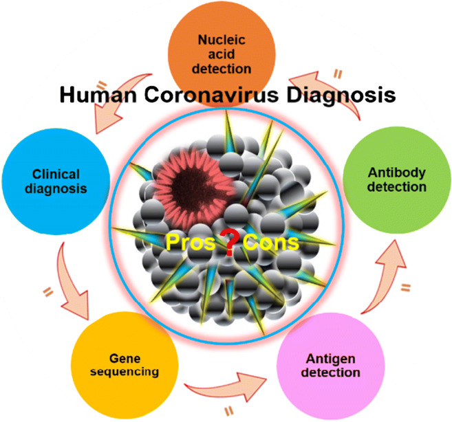
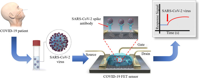
The current global fight against coronavirus disease (COVID-19) to flatten the transmission curve is put forth by the World Health Organization (WHO) as there is no immediate diagnosis or cure for COVID-19 so far. In order to stop the spread, researchers worldwide are working around the clock aiming to develop reliable tools for early diagnosis of severe acute respiratory syndrome (SARS-CoV-2) understanding the infection path and mechanisms. Currently, nucleic acid-based molecular diagnosis (real-time reverse transcription polymerase chain reaction (RT-PCR) test) is considered the gold standard for early diagnosis of SARS-CoV-2. Antibody-based serology detection is ineffective for the purpose of early diagnosis, but a potential tool for serosurveys, providing people with immune certificates for clearance from COVID-19 infection. Meanwhile, there are various blooming methods developed these days. In this review, we summarise different types of coronavirus discovered which can be transmitted between human beings. Methods used for diagnosis of the discovered human coronavirus (SARS, MERS, COVID-19) including nucleic acid detection, gene sequencing, antibody detection, antigen detection, and clinical diagnosis are presented. Their merits, demerits and prospects are discussed which can help the researchers to develop new generation of advanced diagnostic tools for accurate and effective control of human coronavirus transmission in the communities and hospitals.

Keywords: Biosensors; COVID-19; Human coronaviruses; Molecular diagnostics; Serology detection.


Conflict of interest statement

The authors declare that they have no conflict of interest.

Figures

Scheme 1
Scheme 1
The schematics of the content of this review
Fig. 1
Fig. 1
Schematic representation for the promising techniques generally used to detect SARS-CoV-2: (a) RT-PCR method is carried out from the reverse transcription of viral RNA into cDNA which is then amplified using specific primers. The amplification process is confirmed from the fluorescent signal indicating the total number of copies in target sequence. (b) Whole genomic sequencing is a complete gene sequencing method which is complicated and not helpful for urgent and large-scale detection. Generally, the viral RNA is extracted from the specimen going through multiplex amplicon sequencing to identify the nucleic acid [58]. Redrawn with permission. (c) Combined method of LAMP and COVID-19 Penn-RAMP: The Penn-RAMP contains two processes of isothermal amplification, first the RPA was carried out at 37 °C at the cap of the tube and then the LAMP at 63 °C within the tube. The LAMP reaction mixture was added with the LCV dye and the ratio between RPA and LAMP was 1:9. This was incubated at 38 °C for 15–20 min followed by flipping for through mixing moving towards second time incubation in a thermal cycler for 40 min at 63 °C. LCV dye helped by producing dark violet signal in the presence of dsDNA and colourless in the absence of dsDNA. This method was found to have high potential as it reduced false negatives [59]. Re-produced, permission has taken under the CC-BY-NC-ND 4.0 International license)
Fig. 2
Fig. 2
(a) Schematic illustration for the working principle of All-In-One Dual CRISPR-Cas12a (AIOD-CRISPR) assay: demonstrating the stimulation of RPA amplification to reveal the binding sites of Cas 12a-crRNA which turns on the fluorescence on activation of the endonuclease enzyme. (b) Designing and analysing of AIOD-CRISPR assay: The ssDNA-FQ reporter molecule was first marked with the fluorophore-5′6-FAM and a quencher, which was subsequently put through RPA reactive treatments: (i) the reaction system showing direct visualisation of bright fluorescence under LED, blue light, and UV light; (ii) the reactive system number 5 produces highest bright signal due to the presence of smaller DNA sizes cleaved from the reporter molecule; (iii) graph indicating the saturation of highest produced fluorescent signal in 13 min [94]. Reproduced with permission
Fig. 3
Fig. 3
Comparative relation theoretically between different levels of SARS-CoV-2 RNA and antigen, IgM and IgG during the different infection days showing three main phases such as window period, decline phase, and convalescence phase. In the window period, the onset of symptoms takes place within a week of contact with viral source. Secondly, the IgM shows up and the production of IgG takes place until it disappears in 21 days of infection. Thirdly, in the recovery, the IgG remains in the blood. This suggests that the serological examination could be done 3 days after the symptoms or a week after the infection [114]. Reproduced with permission
Fig. 4
Fig. 4
Demonstration of lateral flow immunochromatographic assays for the diagnosis of COVID-19: (a) A CRISPR-CAS12-based assay for detection of viral nucelic acid where the viral RNA was extracted from the swab sample and amplified. Then, as a control, the DETECTR assay was carried out on E, N, and human RNase P gene for 30 min. This was applied for RT-LAMP and Cas12 detection at 62 °C and 37 °C [138] (re-produced, permission has taken under the CC-BY-NC-ND 4.0 International license). (B) RT-LAMP-NBS assay for COVID-19 detection: (i) The amplification mixtures were prepared as LAMP reagents shown in legends, (ii) The RT-Lamp reaction was conducted at 63 °C for 40 min, (iii) The products FIT/biotin-labelled F1ab-LAMP amplicons and np-LAMP amplicons formed [71]. Re-produced, permission has taken under the CC-BY-NC-ND 4.0 International license. (c) Diagrammatic representation of IgM-IgG antibody test strip: The mouse anti-human monoclonal antibodies IgM and IgG were set as the test lines ‘G and M’ on the strip, and the anti-rabbit IgG antibody was used as the control. Then the SARS-CoV-2 antigen was added to the gold colloid to form a conjugate nanoparticle. This was introduced on the pad through spraying on the surface that contains the specific SARS-CoV-2 antibody test lines. Likewise, the gold nanoparticle conjugate of rabbit IgG was also sprayed along to fix with the control line ‘c’ constructing a lateral flow device. The test was performed by running up to 15 μL of specimen and the analysis were determined from the ‘c’ zone by visualisation of red-purple line for the SARS-CoV infection [137]. Re-produced, permission has taken under the CC-BY-NC-ND 4.0 International license. (d) Graphic representation of immunochromatographic strips working under fluroscence: (i) The control line was coated with goat anti-rabbit IgG antibody, the test line was coated with the mouse anti-2019-nCoV NP M1 antibody as shown in the legends. Here the conjugae was the Euphorbium (III) treated with carboxylate that was conjugated to the antibody. (ii) The assembled strip within the plastic cassette containing a well for the sample loading and a testing window. The results through this immunochromatographic technique can be determined when the fluorecent signals are captured through immunofluorecence analyser [4]. Re-produced, permission has taken under the CC-BY-NC-ND 4.0 International license
Fig. 5
Fig. 5
Graphically representation of FET sensor for COVID-19: Firstly, a carbon-based biosensing platform was constructed by functionalising the graphene with viral spike antibody. The functionalisation was done via immobilisation technique, where a coupling agent 1-pyrenebutyric acid N-hydroxysuccinimide ester (PBASE) taking the role of a probe linker. This forms the conjugated graphene sheet capable of detecting 1 fg/mL of viral antigen [152]. Reproduced with permission

References

    1. Tyrrell DA, Bynoe ML. Cultivation of viruses from a high proportion of patients with colds. Lancet. 1966;1(7428):76–77. - PubMed
    1. Callow KA, Parry HF, Sergeant M, Tyrrell DA. The time course of the immune response to experimental coronavirus infection of man. Epidemiol Infect. 1990;105(2):435–446. - PMC - PubMed
    1. Valitutto MT, Aung O, Tun KYN, Vodzak ME, Zimmerman D, Yu JH, et al. Detection of novel coronaviruses in bats in Myanmar. PLoS One. 2020;15(4):e0230802. - PMC - PubMed
    1. Chamings A, Nelson TM, Vibin J, Wille M, Klaassen M, Alexandersen S. Detection and characterisation of coronaviruses in migratory and non-migratory Australian wild birds. Sci Rep. 2018;8(1):5980. - PMC - PubMed
    1. Peiris JSM, Lai ST, Poon LLM, Guan Y, Yam LYC, Lim W, et al. Coronavirus as a possible cause of severe acute respiratory syndrome. Lancet. 2003;361(9366):1319–1325. - PMC - PubMed

MeSH terms

LinkOut - more resources