

An anatomical comparison of the fasciae of the thigh: A macroscopic, microscopic and ultrasound imaging study
- PMID: 33219512
- PMCID: PMC7930759
- DOI: 10.1111/joa.13360
An anatomical comparison of the fasciae of the thigh: A macroscopic, microscopic and ultrasound imaging study
Abstract
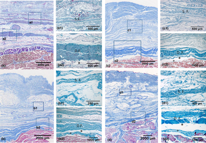
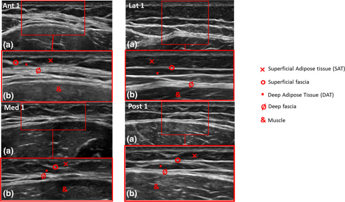
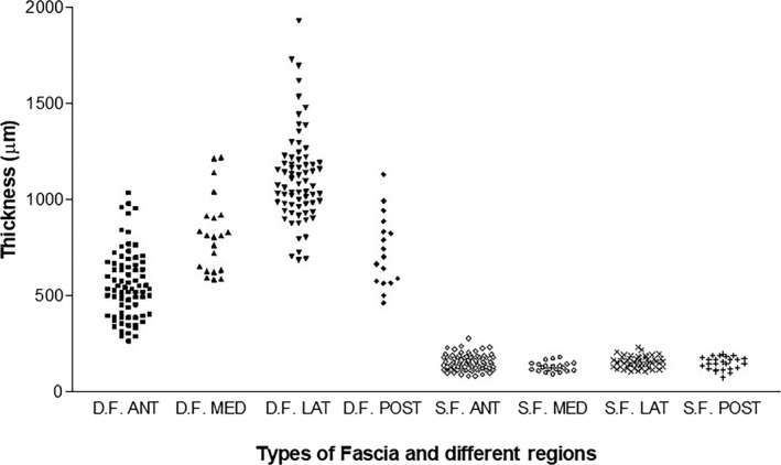
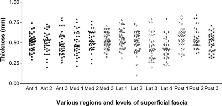
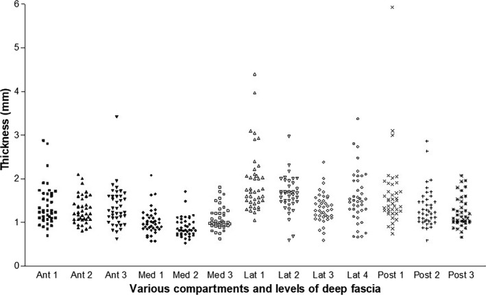
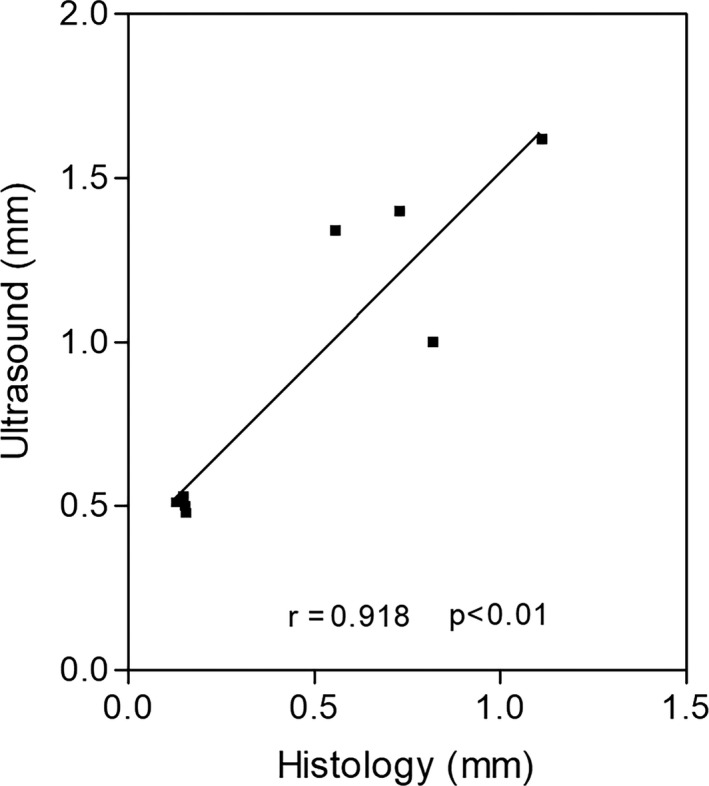
Although the number of Ultrasound (US) imaging studies investigating the fascial layers are becoming more numerous, the majority tend to use different reference points and terminology to describe their findings. The current work set out to compare macroscopic and microscopic data of specimens of the fascial layers of the thigh with US imaging findings. Specimens of the different fascial layers of various regions of the thigh were collected for macroscopic and histological analyses from three fresh cadavers and compared with in vivo US images of the thighs of 20 healthy volunteers. The specimens showed that the subcutaneous tissue of the thigh is made up of three layers: a superficial adipose layer, a membranous layer/superficial fascia, and a deep adipose layer. The deep fascia is composed of an aponeurotic fascia, which envelops all the thigh muscles and is laterally reinforced by the iliotibial tract and an epimysial fascia, which is specific for each muscle. The morphometric measurements of the thickness of the superficial fascia were different (anterior: 153.2 ± 39.3 µm; medial: 128.4 ± 24.7 µm; lateral: 154 ± 28.9 µm; and posterior: 148.8 ± 33.2 µm) as were those of the deep fascia (anterior: 556.8 ± 176.2 µm; medial: 820.4 ± 201 µm; lateral: 1112 ± 237.9 µm; and posterior: 730.4 ± 186.5 µm). The US scans showed a clear picture of the superficial adipose tissue, the superficial fascia, and the deep adipose tissue, as well as the deep fasciae. The epimysial and aponeurotic fasciae of only some topographic areas could be independently identified. The US imaging findings confirmed that the superficial and deep fascia have different thicknesses, and they showed that the US measurements were always larger with respect to those produced by histological analysis (p < 0.001) probably due to shrinkage during the processing. The posterior region (level 1) of the superficial fascia had, for example, a mean thickness of 0.56 ± 0.12 mm at US, while the histological analysis showed that it was 148.8 ± 33.2 µm. Showing a similar pattern, the thickness of the deep fascia was as follows: 1.64 ± 0.85 mm versus 730.4 ± 186.5 µm. Study results have confirmed that US can be considered a valid, non-invasive instrument to evaluate the fascial layers. In any event, there is a clear need for a set of standardised protocols since the thickness of the fascial layers of different parts of the human body varies and the data obtained using inaccurate reference points are not reproducible or comparable. Given the inconsistent terminology used to describe the fascial system, it would also be important to standardise the terminology used to define its parts. The difficulty in distinguishing between the epimysial and aponeurotic/deep fascia can also impede data interpretation.
Keywords: fascia lata; imaging; musculo-skeletal ultrasound; superficial fascia; thickness.
© 2020 Anatomical Society.
Conflict of interest statement
None of the authors has any conflict of interest to disclose.
Figures
Similar articles
-
Distinct displacement of the superficial and deep fascial layers of the iliotibial band during a weight shift task in runners: An exploratory study.J Anat. 2022 Mar;240(3):579-588. doi: 10.1111/joa.13575. Epub 2021 Oct 26. J Anat. 2022. PMID: 34697798 Free PMC article.
-
Elastic Fibres in the subcutaneous tissue: Is there a difference between superficial and muscular fascia? A cadaver study.Skin Res Technol. 2022 Jan;28(1):21-27. doi: 10.1111/srt.13084. Epub 2021 Aug 22. Skin Res Technol. 2022. PMID: 34420234 Free PMC article.
-
Pectoral and femoral fasciae: common aspects and regional specializations.Surg Radiol Anat. 2009 Jan;31(1):35-42. doi: 10.1007/s00276-008-0395-5. Epub 2008 Jul 29. Surg Radiol Anat. 2009. PMID: 18663404
-
Diagnostic ultrasound assessment of deep fascia sliding mobility in vivo: A scoping review - Part 1: Thoracolumbar and abdominal fasciae.J Bodyw Mov Ther. 2021 Jul;27:92-102. doi: 10.1016/j.jbmt.2020.12.027. Epub 2021 Jan 29. J Bodyw Mov Ther. 2021. PMID: 34391319
-
Diagnostic ultrasound assessment of deep fascia sliding mobility in vivo: A scoping review - Part 2: Femoral and crural fasciae.J Bodyw Mov Ther. 2021 Jul;27:84-91. doi: 10.1016/j.jbmt.2021.01.016. Epub 2021 Jan 30. J Bodyw Mov Ther. 2021. PMID: 34391317
Cited by
-
A Closer Look at the Cellular and Molecular Components of the Deep/Muscular Fasciae.Int J Mol Sci. 2021 Jan 30;22(3):1411. doi: 10.3390/ijms22031411. Int J Mol Sci. 2021. PMID: 33573365 Free PMC article. Review.
-
Fascia Lata Alterations in Hip Osteoarthritis: An Observational Cross-Sectional Study.Life (Basel). 2021 Oct 25;11(11):1136. doi: 10.3390/life11111136. Life (Basel). 2021. PMID: 34833012 Free PMC article.
-
Ultrasound Imaging of Crural Fascia and Epimysial Fascia Thicknesses in Basketball Players with Previous Ankle Sprains Versus Healthy Subjects.Diagnostics (Basel). 2021 Jan 26;11(2):177. doi: 10.3390/diagnostics11020177. Diagnostics (Basel). 2021. PMID: 33530583 Free PMC article.
-
Fascial Ultrasound-Guided Injection: Where Do We Really Inject?Cureus. 2025 Feb 11;17(2):e78867. doi: 10.7759/cureus.78867. eCollection 2025 Feb. Cureus. 2025. PMID: 40084316 Free PMC article.
-
Dose-Dependent Pain and Pain Radiation after Chemical Stimulation of the Thoracolumbar Fascia and Multifidus Muscle: A Single-Blinded, Cross-Over Study Revealing a Higher Impact of Fascia Stimulation.Life (Basel). 2022 Feb 25;12(3):340. doi: 10.3390/life12030340. Life (Basel). 2022. PMID: 35330091 Free PMC article.
References
-
- Abrahams, A.C. , Dendooven, A. , van der Veer, J.W. , Wientjes, R. , Toorop, R.J. , Bleys, R.L. et al. (2019) Direct comparison of the thickness of the parietal peritoneum using peritoneal biopsy and ultrasonography of the abdominal wall in patients treated with peritoneal dialysis. Peritoneal Dialysis International, 39(5), 455–464. - PubMed
-
- Abu‐Hijleh, M.F. , Roshier, A.L. , Al‐Shboul, Q. , Dharap, A.S. and Harris, P.F. (2006) The membranous layer of superficial fascia: evidence for its widespread distribution in the body. Surgical and Radiologic Anatomy, 28(6), 606–619. - PubMed
-
- Dauendorffer, J.N. , Bastuji‐Garin, S. , Guéro, S. , Brousse, N. and Fraitag, S. (2009) Shrinkage of skin excision specimens: formalin fixation is not the culprit. British Journal of Dermatology, 160(4), 810–814. - PubMed
-
- Derchi, L. and Rizzatto, G. (2007) Technical requirements. In: Bianchi, S. and Martinoli, C. (Eds.) Ultrasound of the musculoskeletal system. Germany: Springer Verlag, pp. 3–16.
MeSH terms
LinkOut - more resources
Full Text Sources
