

Evidence of Severe Acute Respiratory Syndrome Coronavirus 2 Reinfection After Recovery from Mild Coronavirus Disease 2019
- PMID: 33219681
- PMCID: PMC7890673
- DOI: 10.1093/cid/ciaa1421
Evidence of Severe Acute Respiratory Syndrome Coronavirus 2 Reinfection After Recovery from Mild Coronavirus Disease 2019
Abstract
Background: Positive results from real-time reverse-transcription polymerase chain reaction (rRT-PCR) in recovered patients raise concern that patients who recover from coronavirus disease 2019 (COVID-19) may be at risk of reinfection. Currently, however, evidence that supports reinfection with severe acute respiratory syndrome coronavirus 2 (SARS-CoV-2) has not been reported.
Methods: We conducted whole-genome sequencing of the viral RNA from clinical specimens at the initial infection and at the positive retest from 6 patients who recovered from COVID-19 and retested positive for SARS-CoV-2 via rRT-PCR after recovery. A total of 13 viral RNAs from the patients' respiratory specimens were consecutively obtained, which enabled us to characterize the difference in viral genomes between initial infection and positive retest.
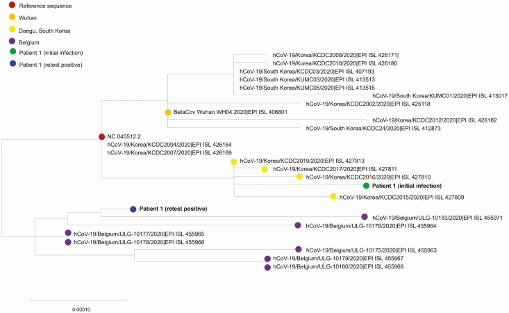
Results: At the time of the positive retest, we were able to acquire a complete genome sequence from patient 1, a 21-year-old previously healthy woman. In this patient, through the phylogenetic analysis, we confirmed that the viral RNA of positive retest was clustered into a subgroup distinct from that of the initial infection, suggesting that there was a reinfection of SARS-CoV-2 with a subtype that was different from that of the primary strain. The spike protein D614G substitution that defines the clade "G" emerged in reinfection, while mutations that characterize the clade "V" (ie, nsp6 L37F and ORF3a G251V) were present at initial infection.
Conclusions: Reinfection with a genetically distinct SARS-CoV-2 strain may occur in an immunocompetent patient shortly after recovery from mild COVID-19. SARS-CoV-2 infection may not confer immunity against a different SARS-CoV-2 strain.
Keywords: COVID-19; SARS-CoV-2; reinfection; whole-genome sequencing.
© The Author(s) 2020. Published by Oxford University Press for the Infectious Diseases Society of America. All rights reserved. For permissions, e-mail: journals.permissions@oup.com.
Figures

Comment in
-
Reinfection With Severe Acute Respiratory Syndrome Coronavirus 2: What Goes Around May Come Back Around.Clin Infect Dis. 2021 Nov 2;73(9):e3009-e3012. doi: 10.1093/cid/ciaa1541. Clin Infect Dis. 2021. PMID: 33035308 Free PMC article. No abstract available.
References
-
- World Health Organization. Coronavirus Disease 2019 (COVID-19): Situation Report–186 (24 July 2020) Available at: https://www.who.int/docs/default-source/coronaviruse/situation-reports/2.... Accessed 11 August 2020.
-
- South Korea reports more recovered coronavirus patients testing positive again Available at:https://www.reuters.com/article/us-health-coronavirus-southkorea/south-k.... Accessed 11 August 2020
-
- Glezen WP, Taber LH, Frank AL, Kasel JA. Risk of primary infection and reinfection with respiratory syncytial virus. Am J Dis Child 1986; 140:543–6. - PubMed
-
- Luo A Positive SARS-Cov-2 test in a woman with COVID-19 at 22 days after hospital discharge: a case report. J Tradit Chin Med Sci 2020. In press.
MeSH terms
Substances
LinkOut - more resources
Full Text Sources
Other Literature Sources
Medical
Miscellaneous
