. 2021 Jan 12;54(1):151-163.e6.
doi: 10.1016/j.immuni.2020.10.014. Epub 2020 Nov 20.

Interleukin-33 Promotes Serotonin Release from Enterochromaffin Cells for Intestinal Homeostasis

Affiliations

Interleukin-33 Promotes Serotonin Release from Enterochromaffin Cells for Intestinal Homeostasis

Zuojia Chen et al. Immunity. .

Abstract

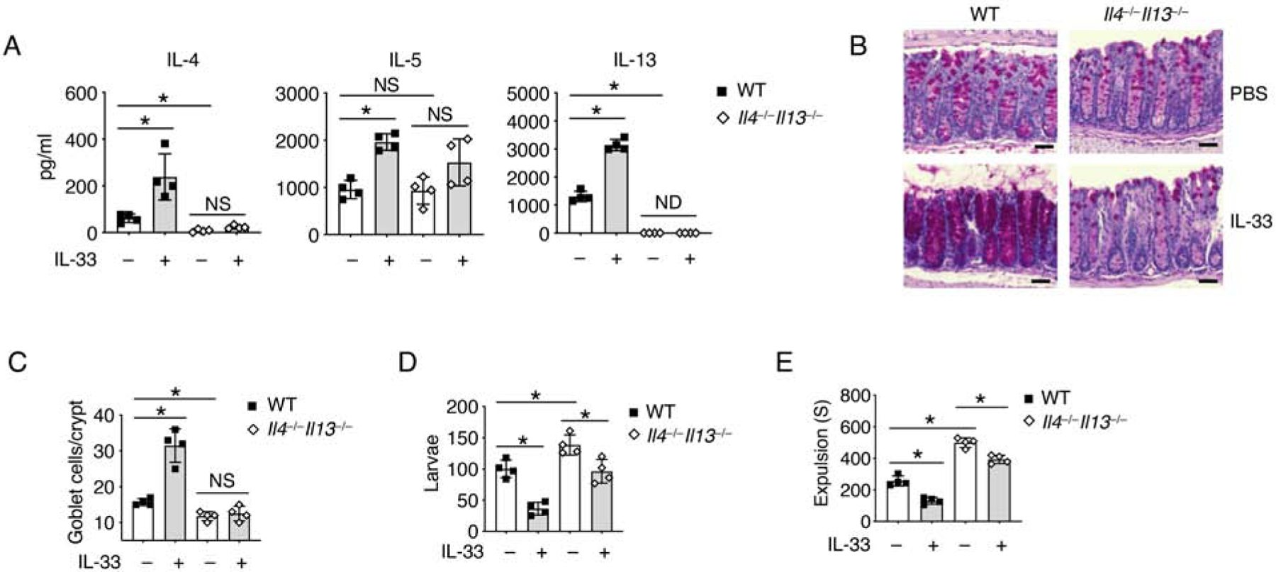
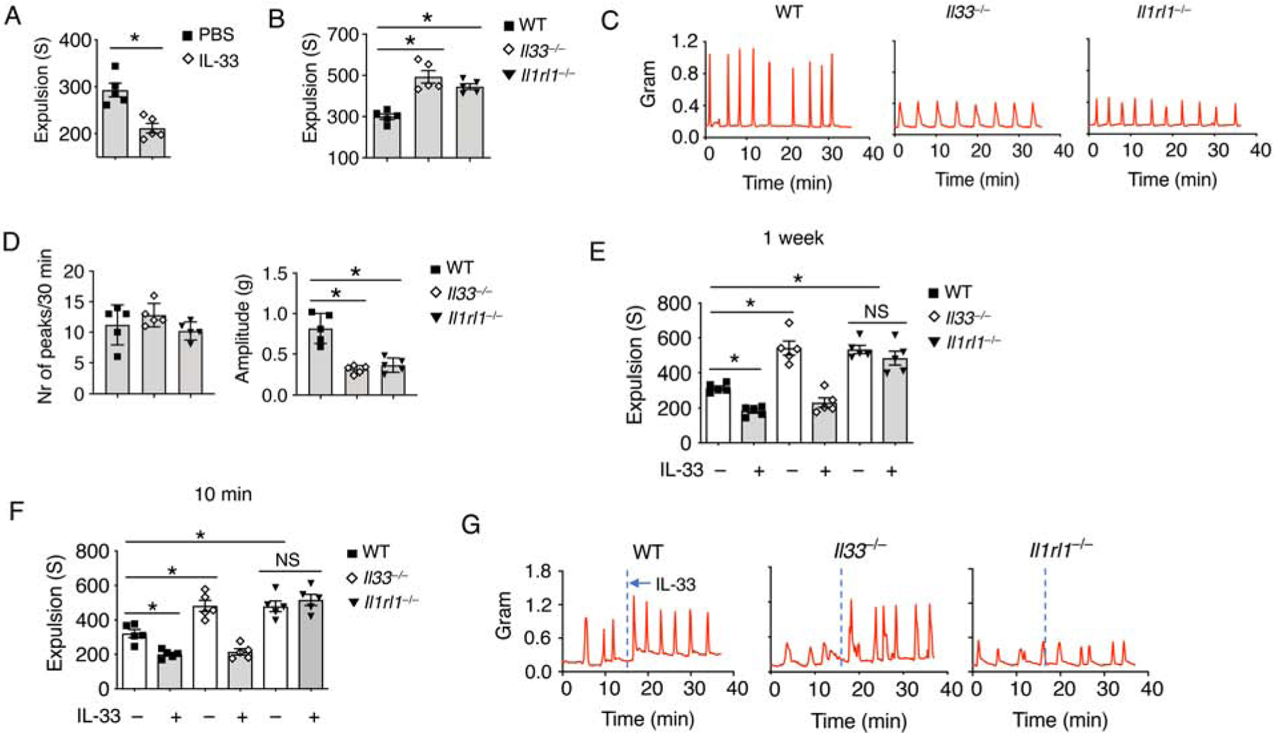
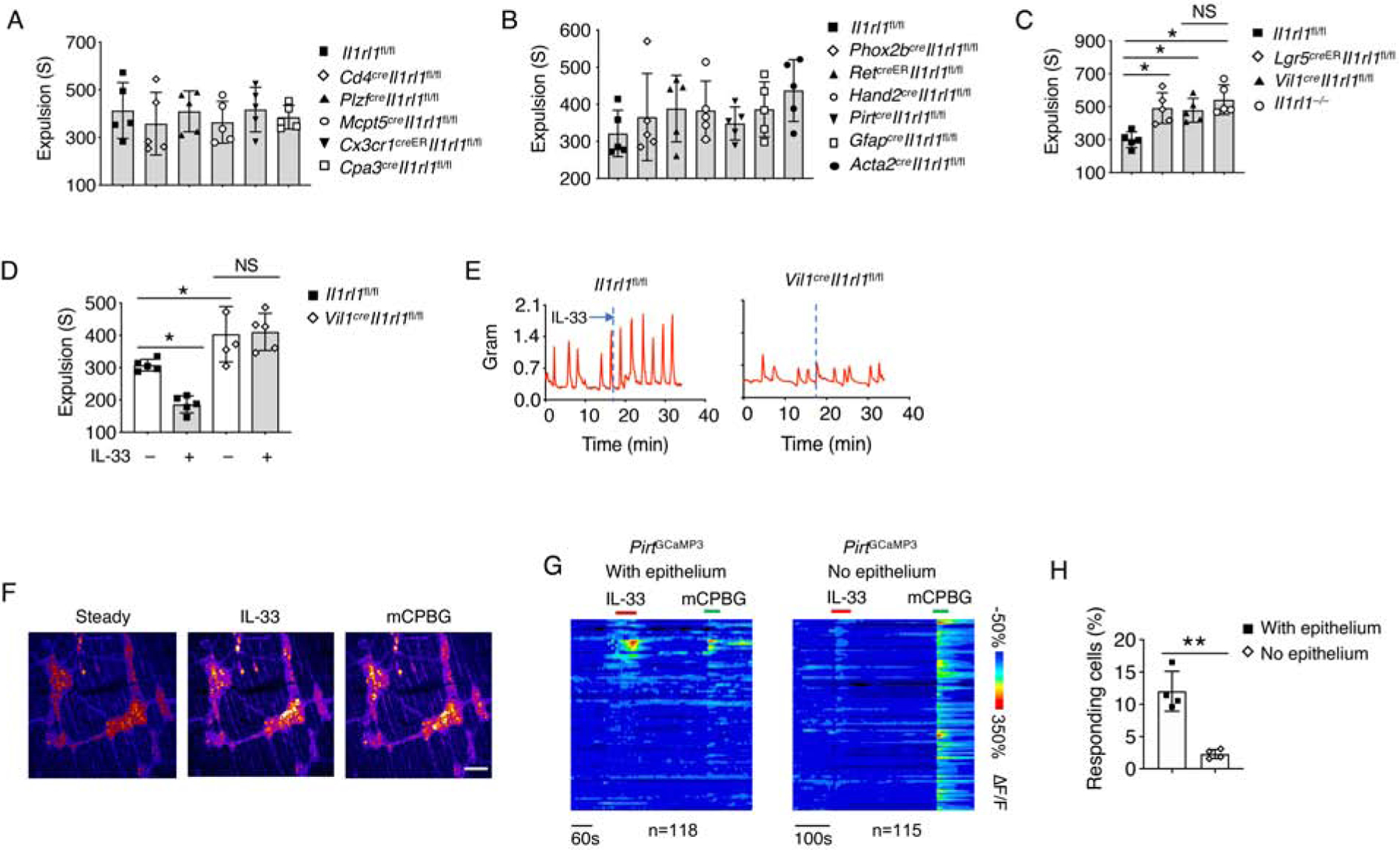
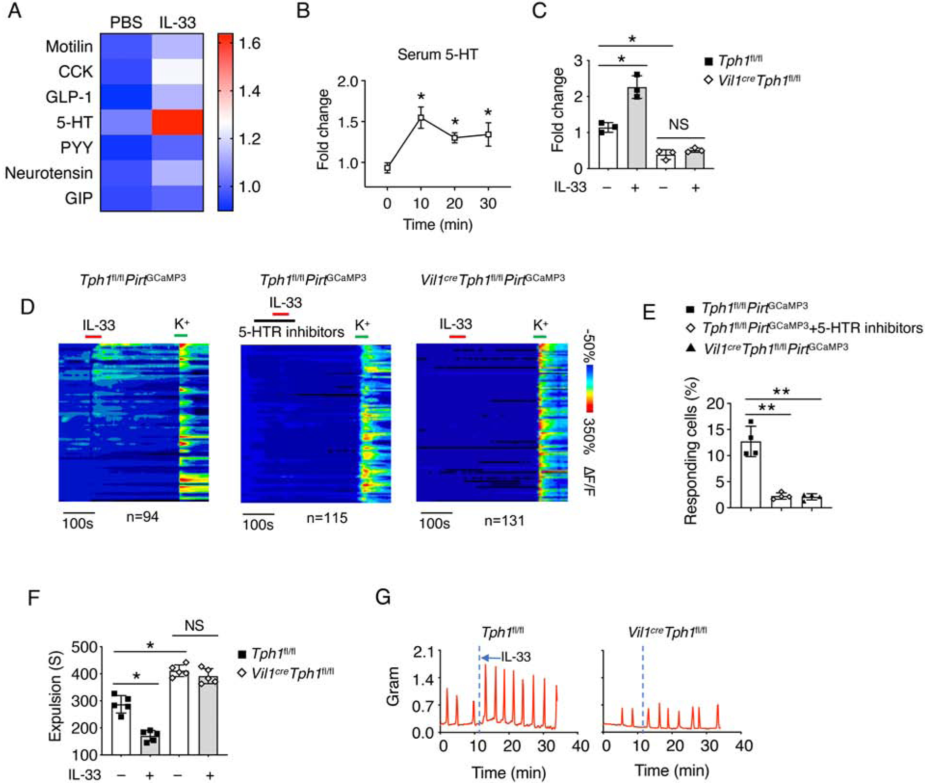
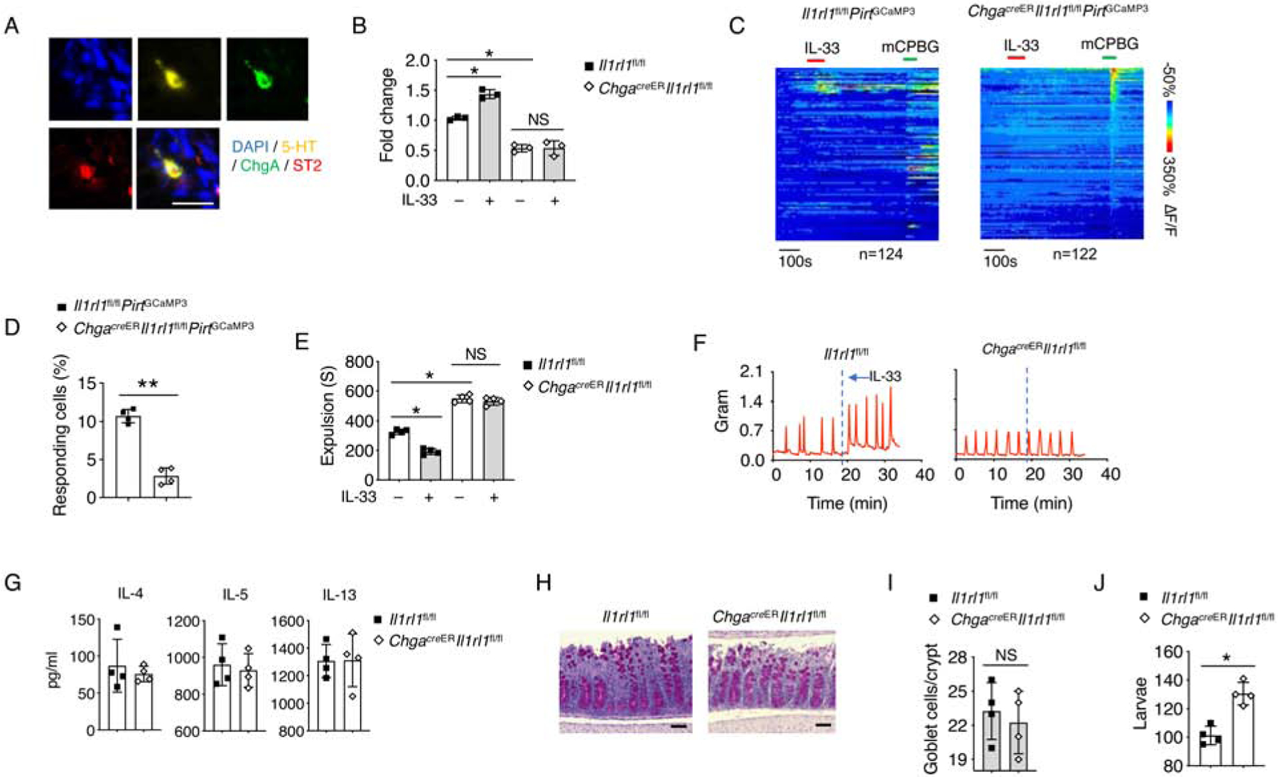
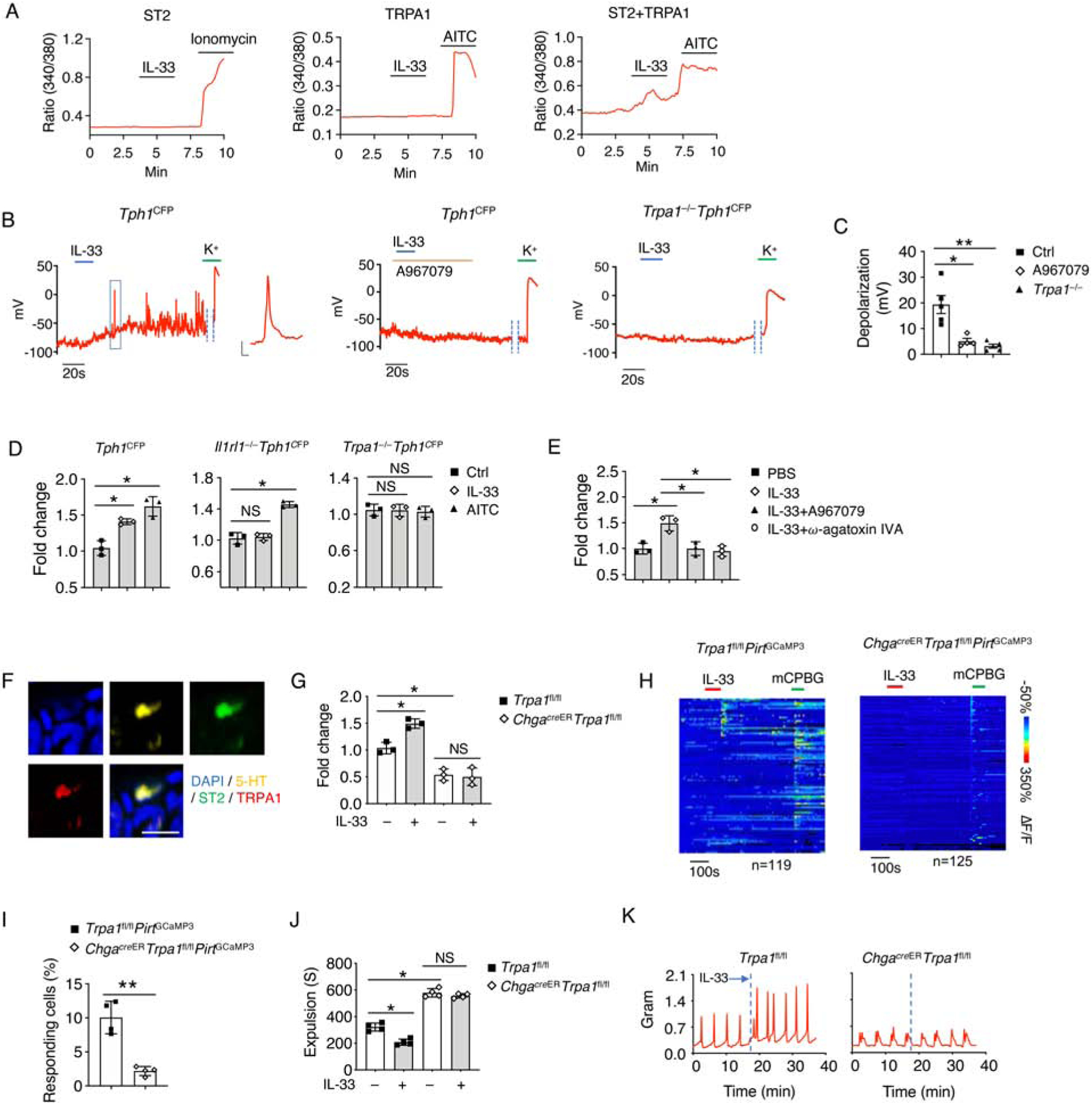
The gastrointestinal tract is known as the largest endocrine organ that encounters and integrates various immune stimulations and neuronal responses due to constant environmental challenges. Enterochromaffin (EC) cells, which function as chemosensors on the gut epithelium, are known to translate environmental cues into serotonin (5-HT) production, contributing to intestinal physiology. However, how immune signals participate in gut sensation and neuroendocrine response remains unclear. Interleukin-33 (IL-33) acts as an alarmin cytokine by alerting the system of potential environmental stresses. We here demonstrate that IL-33 induced instantaneous peristaltic movement and facilitated Trichuris muris expulsion. We found that IL-33 could be sensed by EC cells, inducing release of 5-HT. IL-33-mediated 5-HT release activated enteric neurons, subsequently promoting gut motility. Mechanistically, IL-33 triggered calcium influx via a non-canonical signaling pathway specifically in EC cells to induce 5-HT secretion. Our data establish an immune-neuroendocrine axis in calibrating rapid 5-HT release for intestinal homeostasis.

Keywords: IL-33; PLC-γ1; ST2; TRPA1; enterochromaffin cells; gut motility; helminth clearance; serotonin release.


Conflict of interest statement

Declaration of Interests The authors declare no competing interests.

Figures

Figure 1.
Figure 1.. IL-33 promotes parasite expulsion in a type 2 immune response-independent manner.
(A-E) T. muris eggs were inoculated into WT and Il4−/−Il13−/− mice by oral gavage. The mice were treated with PBS or IL-33 daily from day 7 and sacrificed on 14 day for further test. (A) IL-4, IL-5 and IL-13 protein concentrations in mesenteric lymph nodes (mLN) were assessed by ELISA. (B) PAS staining and (C) Quantification of goblet cells per crypt of colonic tissues. Scale bar, 50 μm. (D) Quantification of larvae in the colons of WT and Il4−/−Il13−/− mice. (E) T. muris infected WT and Il4−/−Il13−/− mice were treated daily with PBS or IL-33 for 7 days. Colonic transit time was assessed by bead expulsion assay. Data are representative of three independent experiments (A-E). NS, not significant; ND, not detected; *p < 0.05 (Student’s t-test, error bars represent SD). Please also see Figure S1.
Figure 2.
Figure 2.. IL-33-ST2 signal regulates gut motility.
(A) WT mice were treated daily with IL-33 or PBS for 7 days. Colonic transit time was assessed by bead expulsion assay. (B) Colonic transit time was measured by bead expulsion assay in WT, Il33−/− and Il1rl1−/− mice. (C) Representative trace and (D) Quantification of colon contraction in WT, Il33−/− and Il1rl1−/− mice. (E) WT, Il33−/− and Il1rl1−/− mice were treated daily with PBS or IL-33 for 7 days. Colonic transit time was assessed by bead expulsion assay at day 8. (F) WT, Il33−/− and Il1rl1−/− mice were treated with PBS or IL-33, and waited for 10 minutes. Colonic transit time measured by bead expulsion assay. (G) Representative trace of IL-33-induced colon contraction in WT, Il33−/− and Il1rl1−/− mice. Data are representative of three independent experiments (C, G) or are pooled from two independent experiments (A, B, D-F). NS, not significant; *p < 0.05 (Student’s t-test, error bars represent SD). Please also see Figure S2.
Figure 3.
Figure 3.. IEC-derived ST2 is required for gut motility.
(A-C) Colonic transit time was assessed by bead expulsion assay in the indicated mouse strains. (D) Il1rl1fl/fl and Vil1creIl1rl1fl/fl mice were treated with PBS or IL-33, and waited for 10 minutes. Colonic transit time was assessed by bead expulsion assay. (E) Representative trace of IL-33-induced colon contraction in Il1rl1fl/fl and Vil1creIl1rl1fl/fl mice. (F) GCaMP3 fluorescence signal of neurons on the myenteric plexus of PirtGCaMP3 mice treated with IL-33 and mCPBG (5HT3R agonist). (G) Time-resolved responses (ΔF/F, color scale) of neurons (one neuron per row) and (H) Quantification of IL-33 responding neurons in the myenteric plexus with or without epithelium. mCPBG was used as a positive control. Data are representative of three independent experiments (E-G) or are pooled from two independent experiments (A-D, H). NS, not significant; *p < 0.05; **p < 0.01 (Student’s t-test, error bars represent SD). Please also see Figure S3.
Figure 4.
Figure 4.. IL-33 induces 5-HT secretion for gut motility.
(A) WT mice were i.p. injected with PBS or IL-33 and waited for 10 minutes. The IEC-derived secretome was assessed from the mice serum. (B) WT mice were treated with IL-33. Mice serum was collected at the indicated time point, and relative 5-HT amounts were assessed by ELISA. (C) Tph1fl/fl and Vil1creTph1fl/fl mice were treated with PBS or IL-33, and waited for 10 minutes. Mice serum was collected, and relative 5-HT amounts were assessed by ELISA. (D) Time-resolved responses (ΔF/F, color scale) of neurons (one neuron per row) and (E) Quantification of IL-33 responding neurons in the myenteric plexus treated with the indicated conditions. K+ was used as a positive control. (F) Tph1fl/fl and Vil1creTph1fl/fl mice were treated with PBS or IL-33, and waited for 10 minutes. Colonic transit time was measured by bead expulsion assay. (G) Representative trace of IL-33-induced colon contraction in Tph1fl/fl and Vil1creTph1fl/fl mice. Data are representative of three independent experiments (A-D, G) or are pooled from two independent experiments (E, F). NS, not significant; *p < 0.05; **p < 0.01 (Student’s t-test, error bars represent SD). Please also see Figure S4.
Figure 5.
Figure 5.. EC cell-derived ST2 responds to IL-33 for 5-HT release.
(A) Representative immunofluorescence staining for 5-HT, chromogranin A (ChgA), ST2 with DAPI in the intestinal tissues of WT mice. Scale bar, 10 μm. (B) Il1rl1fl/fl and ChgacreERIl1rl1fl/fl mice were treated with PBS or IL-33, and waited for 10 minutes. Mice serum was collected, and relative 5-HT amounts were assessed by ELISA (C) Time-resolved responses (ΔF/F, color scale) of neurons (one neuron per row) and (D) Quantification of IL-33 responding neurons in the myenteric plexus. mCPBG (5HT3R agonist) was used as a positive control. (E) Il1rl1fl/fl and ChgacreERIl1rl1fl/fl mice were treated with PBS or IL-33, and waited for 10 minutes. Colonic transit time was measured by bead expulsion assay. (F) Representative trace of IL-33-induced colon contraction in Il1rl1fl/fl and ChgacreERIl1rl1fl/fl mice. (G-J) T. muris eggs were inoculated into Il1rl1fl/fl and ChgacreERIl1rl1fl/fl mice (pre-treated with tamoxifen) by oral gavage and the mice were sacrificed on day 14 for further testing. (G) IL-4, IL-5 and IL-13 protein concentrations from mLN were assessed by ELISA. (H) PAS staining and (I) Quantification of goblet cells per crypt in colonic tissues. Scale bar, 50 μm. (J) Quantification of larvae in the colonic tissues. Data are representative of two independent experiments (A-C, F-J) or are pooled from two independent experiments (D, E). NS, not significant; *p < 0.05; **p < 0.01 (Student’s t-test, error bars represent SD). Please also see Figure S5.
Figure 6.
Figure 6.. TRPA1 is required for IL-33-mediated 5-HT release.
(A) Representative time-lapse Ca2+ imaging traces of IL-33-induced [Ca2+]i response in NIH 3T3 cells overexpressed with ST2 alone; or with TRPA1 alone; or with both ST2 and TRPA1. Ionomycin and AITC were used as positive controls. (B) Representative trace and (C) Quantification of IL-33-induced action potentials were recorded by whole-cell patch-clamp from isolated EC cells of Tph1CFP mice with or without A967079 (TRPA1 antagonist), or isolated EC cells of Trpa1−/−Tph1CFP mice. K+ was used as a positive control. Inset: representative action potential. Scale bar, 20 mV, 10 ms. (D) Relative 5-HT amounts in the cultured supernatant from EC cell-enriched intestinal organoids treated with PBS, IL-33 or AITC (TRPA1 agonist) for 8 hours were assessed by ELISA. (E) Relative 5-HT amounts in the cultured supernatant from EC cell-enriched intestinal organoids treated with the indicated conditions for 8 hours derived from Tph1CFP mice were assessed by ELISA. A967079 (TRPA1 antagonist). ω-agatoxin IVA (Ca2+ channel blocker). (F) Representative immunofluorescence staining for 5-HT, ST2, TRPA1 and DAPI in the colonic tissues of WT mice. Scale bar, 10 μm. (G) Trpa1fl/fl and ChgacreERTrpa1fl/fl mice were treated with PBS or IL-33, and waited for 10 minutes. Mice serum was collected, and relative 5-HT amounts were assessed by ELISA. (H) Time-resolved responses (ΔF/F, color scale) of neurons (one neuron per row) and (I) Quantification of Trpa1fl/flPritGCaMP3 and ChgacreERTrpa1fl/flPritGCaMP3 mice that were treated with the indicated conditions. mCPBG (5HT3R agonist) was used as a positive control. (J) Trpa1fl/fl and ChgacreERTrpa1fl/fl mice were treated with PBS or IL-33, and waited for 10 minutes. Colonic transit time was measured by bead expulsion assay. (K) Representative trace of IL-33-induced colon contraction in Trpa1fl/fl and ChgacreERTrpa1fl/fl mice. Data are representative of three independent experiments (A, B, D-H, J, K) or are pooled from two independent experiments (C, I). NS, not significant; *p < 0.05; **p < 0.01 (Student’s t-test, error bars represent SD). Please also see Figure S6.
Figure 7.
Figure 7.. IL-33 induces PLC-γ1 activation for 5-HT release in EC cells.
(A) Representative trace and (B) Quantification IL-33-induced action potentials were recorded by whole-cell patch-clamp from isolated EC cells of Tph1CFP mice with or without BAPTA. K+ was used as a positive control. Inset: representative action potential. Scale bar, 20 mV, 10 ms. BAPTA (calcium chelator, cell-impermeant). (C) Tph1CFP EC-enriched intestinal organoids were treated with PBS or IL-33 for 10 minutes and fixed. Representative immunofluorescence staining for TPH1-CFP, p-PLC-γ1 and DAPI. Scale bar, 100 μm. (D) Representative trace and (E) Quantification of IL-33-induced action potentials were recorded by whole-cell patch-clamp in isolated EC cells from Tph1CFP mice treated with U-73343 (inactive analog of PLC-γ1 inhibitor U-73122) or U-73122. Inset: representative action potential; K+ was used as a positive control. Scale bar, 20 mV, 10 ms. (F) Relative 5-HT amounts in the cultured supernatant from EC-enriched intestinal organoids treated with the indicated conditions for 8 hours derived from Tph1CFP mice were assessed by ELISA. BAPTA-AM (calcium chelator, cell-permeant). (G) Myd88−/−Tph1CFP EC-enriched intestinal organoids were treated with PBS or IL-33 for 10 minutes and fixed. Representative immunofluorescence staining for TPH1-CFP, p-PLC-γ1 and DAPI. Scale bar, 100 μm. (H) Relative 5-HT amounts in the cultured supernatant from EC-enriched intestinal organoids derived from Tph1CFP and Myd88−/−Tph1CFP mice treated with PBS or IL-33 for 8 hours were assessed by ELISA. (I) Representative immunofluorescence staining for 5-HT, ST2, TRPA1 and DAPI in the healthy human intestinal tissues. Scale bar, 10 μm. (J) Human EC cell-enriched intestinal organoids were treated with PBS or IL-33 for 10 minutes and fixed. Representative immunofluorescence staining for 5-HT, p-PLC-γ1 with DAPI. Scale bar, 100 μm. (K-L) The human EC cell-enriched intestinal organoids were treated with the indicated conditions for 8 hours. Relative 5-HT amounts in the cultured supernatant were assessed by ELISA. Data are representative of three independent experiments (A, C, D, F-L) or are pooled from two independent experiments (B, E). NS, not significant; *p < 0.05; **p < 0.01; ***p < 0.001 (Student’s t-test, error bars represent SD). Please also see Figure S7.

Comment in

References

    1. Alcaino C, Knutson KR, Treichel AJ, Yildiz G, Strege PR, Linden DR, Li JH, Leiter AB, Szurszewski JH, Farrugia G, and Beyder A (2018). A population of gut epithelial enterochromaffin cells is mechanosensitive and requires Piezo2 to convert force into serotonin release. Proceedings of the National Academy of Sciences of the United States of America 115, E7632–E7641. - PMC - PubMed
    1. Antignano F, Mullaly SC, Burrows K, and Zaph C (2011). Trichuris muris infection: a model of type 2 immunity and inflammation in the gut. J Vis Exp. - PMC - PubMed
    1. Baganz NL, and Blakely RD (2013). A dialogue between the immune system and brain, spoken in the language of serotonin. ACS Chem Neurosci 4, 48–63. - PMC - PubMed
    1. Basak O, Beumer J, Wiebrands K, Seno H, van Oudenaarden A, and Clevers H (2017). Induced Quiescence of Lgr5+ Stem Cells in Intestinal Organoids Enables Differentiation of Hormone-Producing Enteroendocrine Cells. Cell Stem Cell 20, 177–190 e174. - PubMed
    1. Bellono NW, Bayrer JR, Leitch DB, Castro J, Zhang C, O’Donnell TA, Brierley SM, Ingraham HA, and Julius D (2017). Enterochromaffin Cells Are Gut Chemosensors that Couple to Sensory Neural Pathways. Cell 170, 185–198 e116. - PMC - PubMed

Publication types