

Rare Case of a Disappearing Pituitary Adenoma During the Coronavirus Disease 2019 (COVID-19) Pandemic
- PMID: 33220478
- PMCID: PMC7673213
- DOI: 10.1016/j.wneu.2020.11.073
Rare Case of a Disappearing Pituitary Adenoma During the Coronavirus Disease 2019 (COVID-19) Pandemic
Abstract
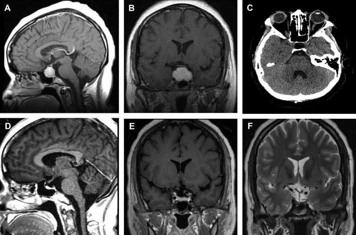
We present a case of a 28-year-old woman with a history of severe headaches and pituitary insufficiency. She was found to have a large, enhancing, sellar mass consistent with a pituitary adenoma. The patient's surgical care was delayed due to the coronavirus disease 2019 (COVID-19) pandemic, and follow-up imaging revealed spontaneous involution of the sellar mass. Spontaneous involution of pituitary masses has been described but not often encountered in clinical practice. This case highlights that follow-up imaging is necessary when scheduling elective surgeries during the COVID-19 pandemic.
Keywords: COVID-19; Pituitary adenoma; Pituitary apoplexy.
Copyright © 2020 Elsevier Inc. All rights reserved.
Figures
References
-
- Bailey P. Pathological report of a case of acromegaly, with special reference to the lesions in the hypophysis cerebri and in the thyroid gland, and a case of hemorrhage into the pituitary. Phila Med J. 1898;1:789–792.
-
- Verrees M., Arafah B.M., Selman W.R. Pituitary tumor apoplexy: characteristics, treatment, and outcomes. Neurosurg Focus. 2004;16:1–7. - PubMed
-
- Micko A.S.G., Wöhrer A., Wolfsberger S., Knosp E. Invasion of the cavernous sinus space in pituitary adenomas: endoscopic verification and its correlation with an MRI-based classification. J Neurosurg. 2015;122:803–811. - PubMed
-
- Villar-Taibo R., Ballesteros-Pomar M.D., Vidal-Casariego A., Álvarez-San Martín R.M., Kyriakos G., Cano-Rodríguez I. Spontaneous remission of acromegaly: apoplexy mimicking meningitis or meningitis as a cause of apoplexy? Arq Bras Endocrinol Metabol. 2014;58:76–80. - PubMed
-
- Robinson D.B., Michaels R.D. Empty sella resulting from the spontaneous resolution of a pituitary macroadenoma. Arch Intern Med. 1992;152:1920–1923. - PubMed
Publication types
MeSH terms
LinkOut - more resources
Full Text Sources
Other Literature Sources
Medical
