

Prognostic Value of Radiotracer-Based Perfusion Imaging in Critical Limb Ischemia Patients Undergoing Lower Extremity Revascularization
- PMID: 33221224
- PMCID: PMC8113329
- DOI: 10.1016/j.jcmg.2020.09.033
Prognostic Value of Radiotracer-Based Perfusion Imaging in Critical Limb Ischemia Patients Undergoing Lower Extremity Revascularization
Abstract
Objectives: The purpose of this study was to evaluate the prognostic value of single-photon emission computed tomography (SPECT)/computed tomography (CT) imaging of angiosome foot perfusion for predicting amputation outcomes in patients with critical limb ischemia (CLI) and diabetes mellitus (DM).
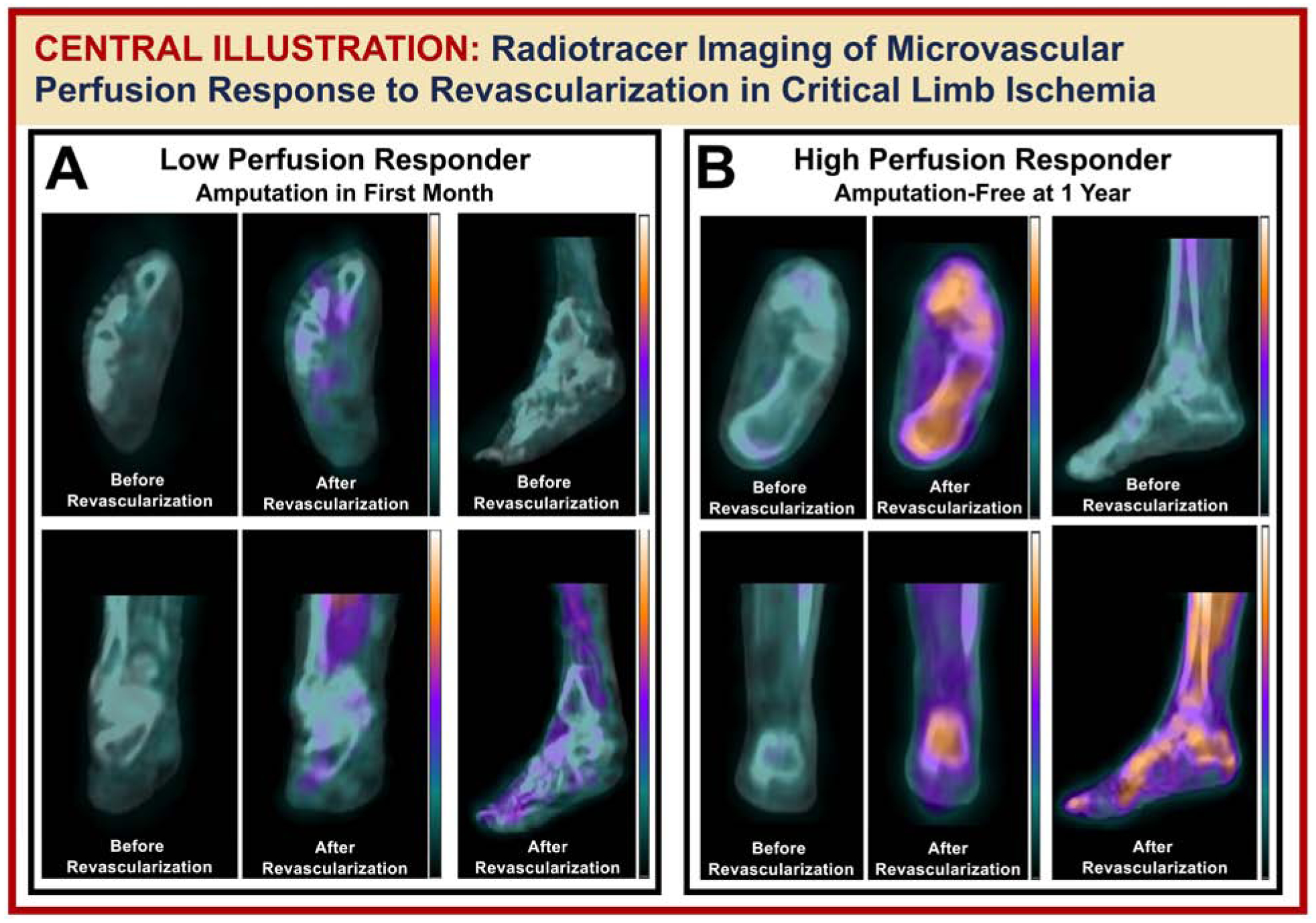
Background: Radiotracer imaging can assess microvascular foot perfusion and identify regional perfusion abnormalities in patients with critical limb ischemia CLI and DM, but the relationship between perfusion response to revascularization and subsequent clinical outcomes has not been evaluated.
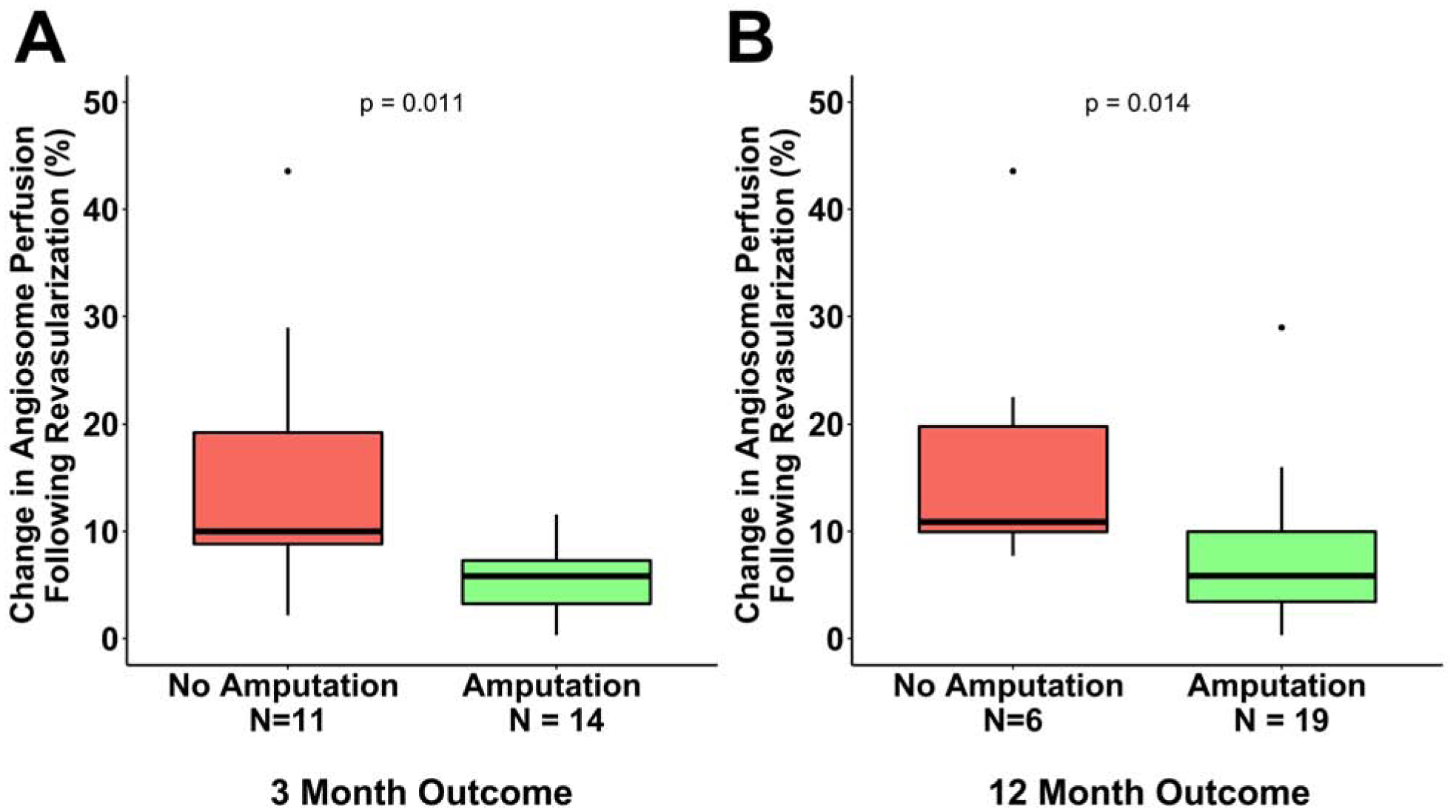
Methods: Patients with CLI, DM, and nonhealing foot ulcers (n = 25) were prospectively enrolled for SPECT/CT perfusion imaging of the feet before and after revascularization. CT images were used to segment angiosomes (i.e., 3-dimensional vascular territories) of the foot. Relative changes in radiotracer uptake after revascularization were evaluated within the ulcerated angiosome. Incidence of amputation was assessed at 3 and 12 months after revascularization.
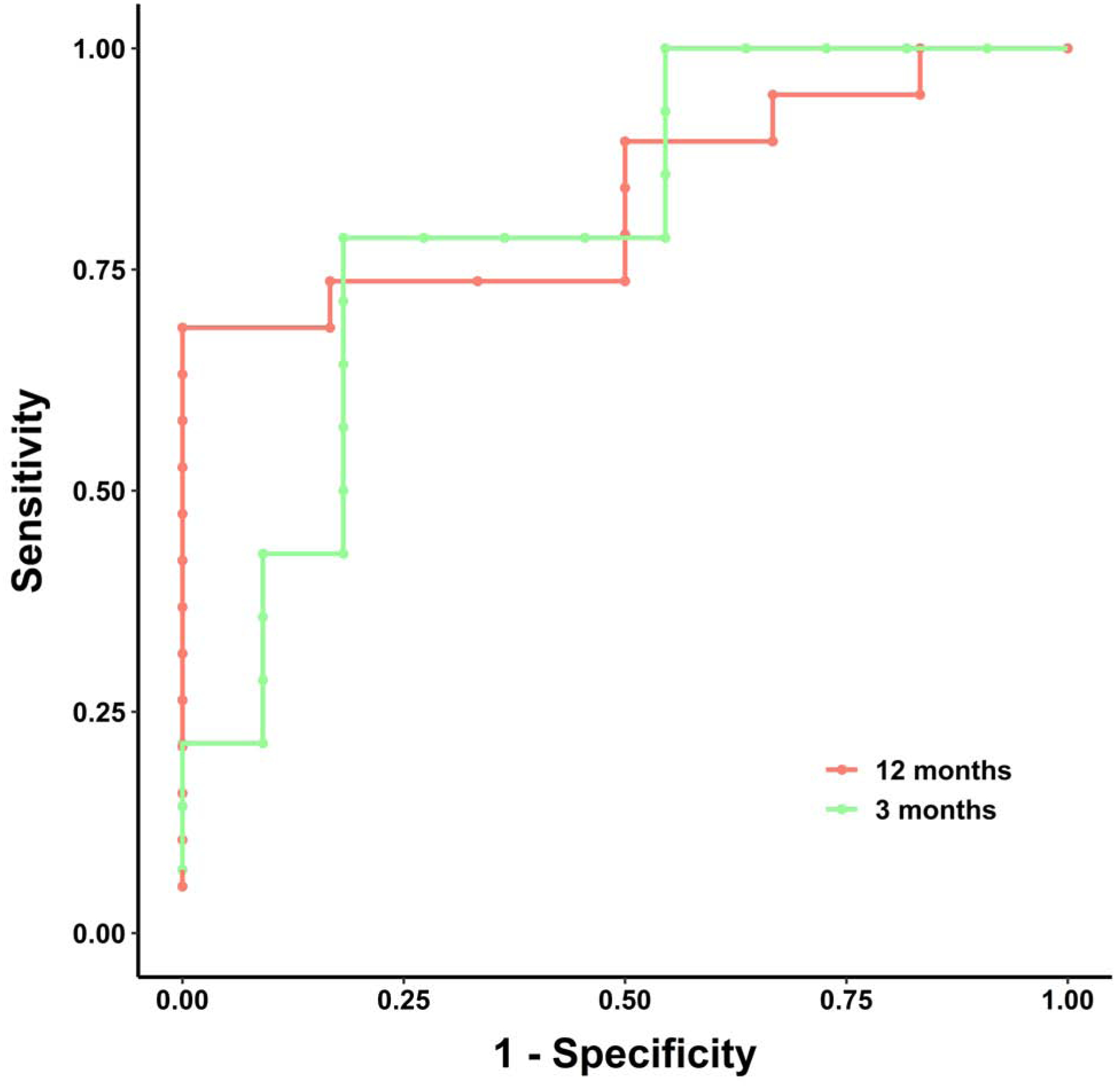
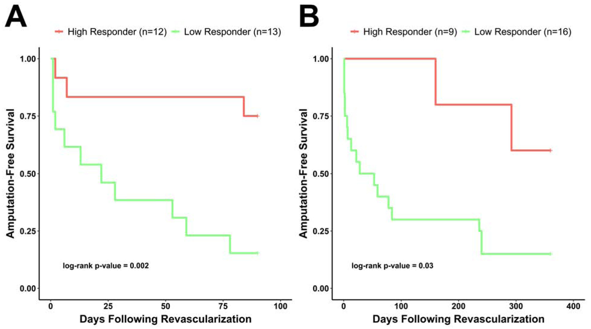
Results: SPECT/CT detected a significantly lower microvascular perfusion response for patients who underwent amputation compared with those who remained amputation free at 3 (p = 0.01) and 12 (p = 0.01) months after revascularization. The cutoff percent change in perfusion for predicting amputation at 3 months was 7.55%, and 11.56% at 12 months. The area under the curve based on the amputation outcome was 0.799 at 3 months and 0.833 at 12 months. The probability of amputation-free survival was significantly higher at 3 (p = 0.002) and 12 months (p = 0.03) for high-perfusion responders than low-perfusion responders to revascularization.
Conclusions: SPECT/CT imaging detects regional perfusion responses to lower extremity revascularization and provides prognostic value in patients with CLI (Radiotracer-Based Perfusion Imaging of Patients With Peripheral Arterial Disease; NCT03622359).
Keywords: critical limb ischemia; diabetes; perfusion imaging; revascularization; single-photon emission computed tomography.
Copyright © 2021 The Authors. Published by Elsevier Inc. All rights reserved.
Conflict of interest statement
Funding Support and Author Disclosures Dr. Stacy has received funding from National Institutes of Health grant R01 HL135103. All other authors have reported that they have no relationships relevant to the content of this paper to disclose.
Figures
Comment in
-
Limb Perfusion Imaging in Peripheral Artery Disease.JACC Cardiovasc Imaging. 2021 Aug;14(8):1625-1627. doi: 10.1016/j.jcmg.2020.10.011. Epub 2020 Nov 18. JACC Cardiovasc Imaging. 2021. PMID: 33221233 No abstract available.
References
-
- Fowkes FG, Rudan D, Rudan I et al. Comparison of global estimates of prevalence and risk factors for peripheral artery disease in 2000 and 2010: a systematic review and analysis. Lancet 2013;382:1329–40. - PubMed
-
- Writing Group M, Mozaffarian D, Benjamin EJ et al. Heart Disease and Stroke Statistics-2016 Update: A Report From the American Heart Association. Circulation 2016;133:e38–360. - PubMed
-
- Farber A, Eberhardt RT. The Current State of Critical Limb Ischemia: A Systematic Review. JAMA Surg 2016;151:1070–1077. - PubMed
Publication types
MeSH terms
Associated data
Grants and funding
LinkOut - more resources
Full Text Sources
Medical
Research Materials
Miscellaneous
