. 2021 Feb:143:115764.
doi: 10.1016/j.bone.2020.115764. Epub 2020 Nov 20.

Diabetes impairs periosteal progenitor regenerative potential

Affiliations

Diabetes impairs periosteal progenitor regenerative potential

Laura Doherty et al. Bone. 2021 Feb.

Abstract

Diabetics are at increased risk for fracture, and experience severely impaired skeletal healing characterized by delayed union or nonunion of the bone. The periosteum harbors osteochondral progenitors that can differentiate into chondrocytes and osteoblasts, and this connective tissue layer is required for efficient fracture healing. While bone marrow-derived stromal cells have been studied extensively in the context of diabetic skeletal repair and osteogenesis, the effect of diabetes on the periosteum and its ability to contribute to bone regeneration has not yet been explicitly evaluated. Within this study, we utilized an established murine model of type I diabetes to evaluate periosteal cell differentiation capacity, proliferation, and availability under the effect of a diabetic environment. Periosteal cells from diabetic mice were deficient in osteogenic differentiation ability in vitro, and diabetic mice had reduced periosteal populations of mesenchymal progenitors with a corresponding reduction in proliferation capacity following injury. Additionally, fracture callus mineralization and mature osteoblast activity during periosteum-mediated healing was impaired in diabetic mice compared to controls. We propose that the effect of diabetes on periosteal progenitors and their ability to aid in skeletal repair directly impairs fracture healing.

Keywords: Advanced glycation end products (AGEs); Diabetic fracture healing; Mesenchymal progenitors; Periosteum; Skeletal repair.


Conflict of interest statement

Declaration of Interests

The authors report no conflict of interest.

Figures

Fig. 1.
Fig. 1.
(A) Schematic of in vitro diabetic environment osteogenic assays. Periosteal cells were isolated from tibias and femurs and cultured to confluence. Cells were replated and exposed to 4-hydroxytamoxifen (1 mM) to induce αSMA-driven Ai9 expression, followed by osteogenic differentiation for 21 days as described. For treatment with both high glucose + AGEs, cells were pretreated for one day prior to osteogenesis with 2 μg/mL AGEs. (B) Cells were stained with alizarin red S to visualize calcium deposits. Insets=10x (C) RNA was isolated from these cells after 21 days of osteogenic differentiation, and analyzed for expression levels of the late osteogenic gene Ocn. N=3 **p<0.005, ***p<0.0005 (D) After 21 days of osteogenic differentiation, cells were analyzed for GFP fluorescence (indicating mature osteoblastic colonies) and αSMA-driven Ai9 (indicative of undifferentiated cells). Scale bar = 250 μm. Images are representative of three separate experiments.
Fig. 2.
Fig. 2.
(A) Schematic of STZ-induced mouse model. Mice were injected with vehicle or a high dose of STZ, and four weeks, later, fasting blood glucose measurements and weight were recorded to confirm diabetic status. Periosteal cells were isolated from tibias and femurs for in vitro assays. (B) Primary periosteal cells were cultured to confluence in basal media. Cells were isolated at confluence, or seeded at the same density in 12-well plates and induced to differentiate toward the osteoblast lineage for 7 or 21 days. (C) At confluence, periosteal cells were isolated and cultured, then analyzed for relative expression of Sp7 and Runx2 via qRT-PCR. N=3 *p<0.05 (D) After 7 days of osteogenic differentiation, cells were analyzed for expression levels of early osteogenic genes and transcription factors, including Alpl, Sp7, and Runx2. N=3 *p<0.05, **p<0.005 (E) After 21 days of osteogenic differentiation, cells were analyzed for expression levels of late osteogenic genes including Ocn and type I collagen (Col1a1). N=3 *p<0.05 (F) After 21 days of osteogenic differentiation, plates were analyzed for GFP+ mature osteoblastic colonies. Images representative of three separate experiments. Scale bar = 250 μm (D)
Fig. 3.
Fig. 3.
(A) Schematic of flow cytometry-based approach to assess periosteal proliferation before and after a fracture insult. Mice were injected with vehicle or a high dose of STZ, and four weeks, later, fasting blood glucose measurements and weight were recorded to confirm diabetic status. Mice underwent femoral fracture and were injected with EdU one day prior to sacrifice. Periosteal cells were isolated from tibias and femurs of either intact tibias and femurs, or from fractured femurs three days after injury. Cells were stained for cell surface markers and analyzed by flow cytometry using the gating strategy in (B). Periosteal cells from intact bones and contralateral fractured femurs were analyzed for the total proliferating (EdU+) cell populations (C) as well as the Sca1+EdU+ proliferating population (D). N=4 *p<0.05
Fig. 4.
Fig. 4.
(A) Schematic of flow cytometry-based approach. Mice were injected with vehicle or a high dose of STZ, and four weeks, later, fasting blood glucose measurements and weight were recorded to confirm diabetic status. Periosteal cells were isolated from tibias and femurs of either intact tibias and femurs, or from fractured femurs three days after injury. Cells were stained for cell surface markers and analyzed by flow cytometry as shown in (B). Periosteal cells from intact femurs (C) in diabetic mice and controls were analyzed for a specific population of mesenchymal progenitors, as defined by LinSca1+CD105+ cells. (D) Periosteal cells isolated from the fracture site in diabetic mice and controls were analyzed for a population of mesenchymal progenitors, as defined by LinSca1+CD105+ cells. (D) Periosteal cells isolated from the fracture site in diabetic mice and controls were analyzed for a population of bona fide mouse skeletal stem cells defined as CD90-CD200+ cells from the parent Lin-CD51+Ly51-CD105- population (E) and the downstream bone, cartilage, and stromal progenitors (BCSPs), defined as CD90-CD105- cells from the parent Lin-CD51+Ly51- population (F). N=4 *p<0.05
Fig. 5.
Fig. 5.
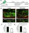
(A) Schematic of the fracture methodology using the STZ-induced Col2.3GFP/SMA9 mouse model. Mice were injected with vehicle or a high dose of STZ, and four weeks, later,, fasting blood glucose measurements and weight were recorded to confirm diabetic status. Mice underwent closed, stabilized femoral fracture. 14 days after fracture, limbs were isolated for histological analysis. (B) Microscopy images demonstrating the use of a Col2.3-driven GFP expression to indirectly visualize osteoblastic mineralizing activity within the callus. Insets show an enlarged view of newly formed bone within the fracture callus. CB=cortical bone, SM=skeletal muscle. Inset = 10x (C) GFP fluorescence intensity of the entire callus as defined in Fig. S1A was quantified 14 days post-fracture. (D) Areas of the callus specific to periosteal-mediated bone formation (Fig. S1B) were analyzed for the GFP expression to indirectly visualize osteoblastic mineralizing activity, and this fluorescence intensity specific to periosteal new bone was quantified 14 days post-fracture. N=3–4 individual mice, *p<0.05

Similar articles

Cited by

References

    1. Yan W, Li X. Impact of diabetes and its treatments on skeletal diseases. Frontiers of Medicine. 2013;7(1):81–90. doi:10.1007/s11684-013-0243-9 - DOI - PubMed
    1. Engelgau MM, Geiss LS, Saaddine JB, Boyle JP, Benjamin SM, Gregg EW, Tierney EF, Rios-Burrows N, Mokdad AH, Ford ES, et al. The evolving diabetes burden in the United States. Annals of Internal Medicine. 2004;140(11):945–950. doi:10.7326/0003-4819-140-11-200406010-00035 - DOI - PubMed
    1. American Diabetes Association. Economic Costs of Diabetes in the U.S. in 2017. Diabetes Care. 2018;41(5):917–928. doi:10.2337/dci18-0007 - DOI - PMC - PubMed
    1. Yamagishi S Role of Advanced Glycation End Products (AGEs) in Osteoporosis in Diabetes. Current Drug Targets. 2011;12(14):2096–2102. doi:10.2174/138945011798829456 - DOI - PubMed
    1. Ramasamy R, Yan SF, Schmidt AM. Receptor for AGE (RAGE): signaling mechanisms in the pathogenesis of diabetes and its complications: RAGE, signal transduction, and diabetes. Annals of the New York Academy of Sciences. 2011;1243(1):88–102. doi:10.1111/j.1749-6632.2011.06320.x - DOI - PMC - PubMed

Publication types

LinkOut - more resources