Review
. 2021 Jun;42(2):305-322.
doi: 10.1007/s10974-020-09591-6. Epub 2020 Nov 22.

T-tubule remodeling in human hypertrophic cardiomyopathy

Affiliations
Review

T-tubule remodeling in human hypertrophic cardiomyopathy

Giulia Vitale et al. J Muscle Res Cell Motil. 2021 Jun.

Abstract

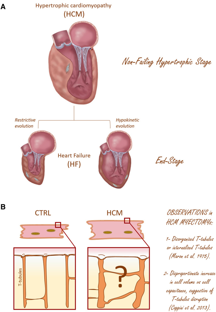
The highly organized transverse T-tubule membrane system represents the ultrastructural substrate for excitation-contraction coupling in ventricular myocytes. While the architecture and function of T-tubules have been well described in animal models, there is limited morpho-functional data on T-tubules in human myocardium. Hypertrophic cardiomyopathy (HCM) is a primary disease of the heart muscle, characterized by different clinical presentations at the various stages of its progression. Most HCM patients, indeed, show a compensated hypertrophic disease ("non-failing hypertrophic phase"), with preserved left ventricular function, and only a small subset of individuals evolves into heart failure ("end stage HCM"). In terms of T-tubule remodeling, the "end-stage" disease does not differ from other forms of heart failure. In this review we aim to recapitulate the main structural features of T-tubules during the "non-failing hypertrophic stage" of human HCM by revisiting data obtained from human myectomy samples. Moreover, by comparing pathological changes observed in myectomy samples with those introduced by acute (experimentally induced) detubulation, we discuss the role of T-tubular disruption as a part of the complex excitation-contraction coupling remodeling process that occurs during disease progression. Lastly, we highlight how T-tubule morpho-functional changes may be related to patient genotype and we discuss the possibility of a primitive remodeling of the T-tubule system in rare HCM forms associated with genes coding for proteins implicated in T-tubule structural integrity, formation and maintenance.

Keywords: Excitation–contraction coupling; Hypertrophic cardiomyopathy; T-tubules.


Conflict of interest statement

The authors declare that they have no conflict of interest.

Figures

Fig. 1
Fig. 1
T-tubule organization in human and rodent ventricular myocytes. A Confocal images of the T-tubule system in tissue sections from human ventricle (top, left) and rat ventricle (top, right), labeled with wheat germ agglutinin (WGA) and lipophilic membrane indicator FM4-64, respectively. Three dimensional reconstructions of single cardiomyocytes from human and rat ventricle loaded with WGA are shown in the lower panels. Scale bars: 20 µm. B WGA labelling of T-tubules in normal and failing human ventricular myocytes. The top row shows images from normal cells in longitudinal and transverse sections (a-d, left to right) and corresponding images from diseased tissue is shown in the lower two rows. (a) Longitudinal sections of normal tissue shows uniformly spaced T-tubules. Occasional axial elements can also be seen. (b) A magnified view of the region shown by the box in a. (c) Normal myocyte in transverse section. A radial ‘‘spoke-like’’ organization of T-tubules is apparent. (d) Enlarged view of the region shown by the box in c. (e, i, k) Longitudinal sections from three different cells from failing heart, demonstrating the range of T-tubular morphologies found in HF with corresponding (f, j, l) magnified views. Note that while the enlarged view in l appears relatively normal, other regions with the same cell (k) are clearly abnormal. (g) Transverse section showing that, while the general direction of diseased tubules is radial, tubules are more disorganized. (h) Magnified view of the region shown by the box in g. Images are projections of 5 slices with z depth of 1 mm. Scale bars in overview images are 10 mm and in close up images 2 mm. HF, heart failure. Reproduced from Manfra et al. (2017) and Crossman et al. (2011)
Fig. 2
Fig. 2
(previous page). HCM: clinical staging and cardiomyocytes remodeling. A Stages of hypertrophic cardiomyopathy from the clinical standpoint. The pathogenic HCM mutations initiate a life-long remodeling process within the myocardium which presents with distinct clinical disease stages. The “Non-failing hypertrophic stage” which is characterized by an hypertrophied and hyperdinamic LV (with an ejection fraction > 65%). About 75% of HCM patients belong to this class. Importantly, during this stage patients may undergo cardiac surgery, named “myectomy”, to relieve LV outflow obstruction, thus giving the possibility to collect samples for biophysical studies. The “end-stage” condition instead is reached by a small subset of patients (5%). This latter condition is characterized by severe functional deterioration of the LV (defined by an LVEF < 50%), clinical decompensation and terminal HF. Sometimes patients are implanted with a contraction assist device (LVAD) or heart transplanted; these events represent another source of myocardial samples. Modified from Coppini et al. (2014). B HCM versus Normal Heart. In normal heart, T-tubules are periodically located at the level of Z-lines, and are rich of contact points with the SR, forming calcium release units (CRUs). This organization is crucial in ensuring a homogeneous Ca2+-release throughout the cell, thus allowing synchronous myofilaments contraction. In HCM hearts, cardiomyocytes appeared hypertrophied and a structural remodeling of the T-tubular network may be present but data on myoctomy samples are scarce and difficult to collect. Different cell types were coexisting in the same diseased hearts and were classified as hypertrophied but non-degenerated cells or cells with evidence of mild to severe degeneration (Maron et al. 1975a, b)
Fig. 3
Fig. 3
Gene mutations associated to HCM. Cartoon depicting the sarcomeres and the associated T-tubule sarcoplasmic reticulum structures. About 35–60% of patients with HCM are heterozygous for missense or truncating mutations in genes encoding sarcomeric proteins, with the most commonly involved being MYBPC3 (cardiac myosin-binding protein-C), MYH7 (β-myosin heavy-chain), and TNNT2 (Troponin T) or TPM1 (Tropomyosin). Rare forms of HCM (prevalence < 1%) are those associated to other genes that are listed on the right panel. Among them, additional sarcomere proteins and Z-line proteins, e.g. TnC, Troponin C; TnI, Troponin I, LC, light chain; TTN, Titin, OBSCN, Obscurine; or proteins involved in E–C coupling and muscle regulation/development (JPH2, Junctophillin 2; CAV3, Caveolin-3; CSRP3, Muscle LIM Protein; NEXN, Nexilin; TCAP, Telethonin)
Fig. 4
Fig. 4
T-tubule remodeling in human HCM myectomies. A Left: Representative images of a control (top) and an HCM (bottom) cardiomyocyte, showing cell hypertrophy in HCM. Right: Surface/volume ratio in HCM and control cardiomyocytes; surface is derived from cell capacitance, volume estimated from cell area. Data from 64 cells (14 patients). From Coppini et al. (2018). B The density of T-tubules is markedly low in HCM cardiomyocytes. Representative confocal images of single cardiomyocytes. Each cell derives from a different HCM patient sample (ID of the patient is indicated next to the cell in each respective image). Cells were stained with Di-3ANEPPDHQ (Thermo-Fisher) and imaged with a Leica Confocal microscope using the 488 nm laser line. Sections were taken at mid cell. While the outer sarcolemma is well stained in all myocytes, T-tubules are barely visible in most of them and some cells are completely devoid of T-tubules. White bars equal 10 μm. Modified from Ferrantini et al. (2018). C Loss of transverse tubules and functionality of axial components in human HCM cardiomyocytes. Two photon fluorescence image of one Di-4-AN(F)EPPTEA labelled HCM trabecula from the left ventricle. The lines mark the probed sarcolemmal regions: surface sarcolemma (SS) in red and axial tubules (AT) in green. White bars equal 10 μm
Fig. 5
Fig. 5
Alterations of T-tubules in mouse models of HCM. A Defects of T-tubules electrical activity and local calcium release in cTnT Δ160E mouse model. Left: two-photon fluorescence (TPF) image of a stained cTnT Δ160E and a WT ventricular myocyte: sarcolemma in magenta (di-4-AN(F)EPPTEA) and [Ca2+]i in green (GFP-certified Fluoforte). Scale bar in white: 5 μm. Right: representative normalized fluorescence traces (ΔF/F0) of SS and two T-tubules (TTi) recorded in WT and cTnT Δ160E cardiomyocyte (average of ten subsequent trials). Membrane potential in magenta, [Ca2+]i in green. AP elicited at 200 ms (black arrowheads). Middle: (top) Columns showing the percentage of electrically failing T-tubules in WT and cTnT Δ160E myocytes. Data from 101 WT and 66 cTnT Δ160E T-tubules (Student’s t-test ***p b 0.001). (bottom) Superposition of fluorescence Ca2+ traces (ΔF/F0) of electrically coupled (AP+, dark green) and uncoupled (AP−, green) T-tubules reported above. The two grey arrows pinpoint Ca2+ transients TTP of the traces. Electrical trigger provided at 200 ms (black arrowhead). (right) Columns showing time-to-peak (TTP) mean values of Ca2+ release measured in cTnT Δ160E cells with respect to WT. Ca2+ transient kinetics is reported by separately analysing the two populations of T-tubules (AP+ and AP−). Data reported as mean ± SEM from 101 WT T-tubules, 65 AP+, and 15 AP− (n = 28 WT and 17 cTnT Δ160E; N = 10WT and 7 cTnT Δ160E). Student’s t-test **p b 0.01, ***p b 0.001. Modified from Crocini et al. (2016). B Left: Representative confocal images from isolated LV cardiomyocytes stained with di-3-aneppdhq from WT, R92Q, R92L, ∆160 and E163R hearts. Horizontal bar equals 10 µm. Right: Columns showing T-tubule Power, as calculated using the TTorg ImageJ plugin, and non-transverse components in cardiomyocytes from the five cohorts of mice. Means ± S.E. Modified Statistics: One-way ANOVA with Tukey correction.*P < 0.05

Similar articles

Cited by

References

    1. Agrawal PB, et al. SPEG interacts with myotubularin, and its deficiency causes centronuclear myopathy with dilated cardiomyopathy. Am J Hum Genet. 2014;95:218–226. doi: 10.1016/j.ajhg.2014.07.004. - DOI - PMC - PubMed
    1. Al-Qusairi L, et al. T-tubule disorganization and defective excitation-contraction coupling in muscle fibers lacking myotubularin lipid phosphatase. Proc Natl Acad Sci USA. 2009;106:18763–18768. doi: 10.1073/pnas.0900705106. - DOI - PMC - PubMed
    1. Arber S, et al. MLP-deficient mice exhibit a disruption of cardiac cytoarchitectural organization dilated cardiomyopathy, and heart failure. Cell. 1997;88:393–403. doi: 10.1016/S0092-8674(00)81878-4. - DOI - PubMed
    1. Ariga R, et al. Identification of myocardial disarray in patients with hypertrophic cardiomyopathy and ventricular arrhythmias. J Am Coll Cardiol. 2019;73:2493–2502. doi: 10.1016/j.jacc.2019.02.065. - DOI - PMC - PubMed
    1. Beavers DL, Landstrom AP, Chiang DY, Wehrens XHT. Emerging roles of junctophilin-2 in the heart and implications for cardiac diseases. Cardiovasc Res. 2014;103:198–205. doi: 10.1093/cvr/cvu151. - DOI - PMC - PubMed

Publication types

LinkOut - more resources