An official website of the United States government
The .gov means it’s official.
Federal government websites often end in .gov or .mil. Before
sharing sensitive information, make sure you’re on a federal
government site.
The site is secure.
The https:// ensures that you are connecting to the
official website and that any information you provide is encrypted
and transmitted securely.
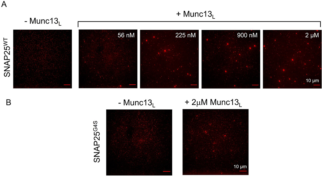
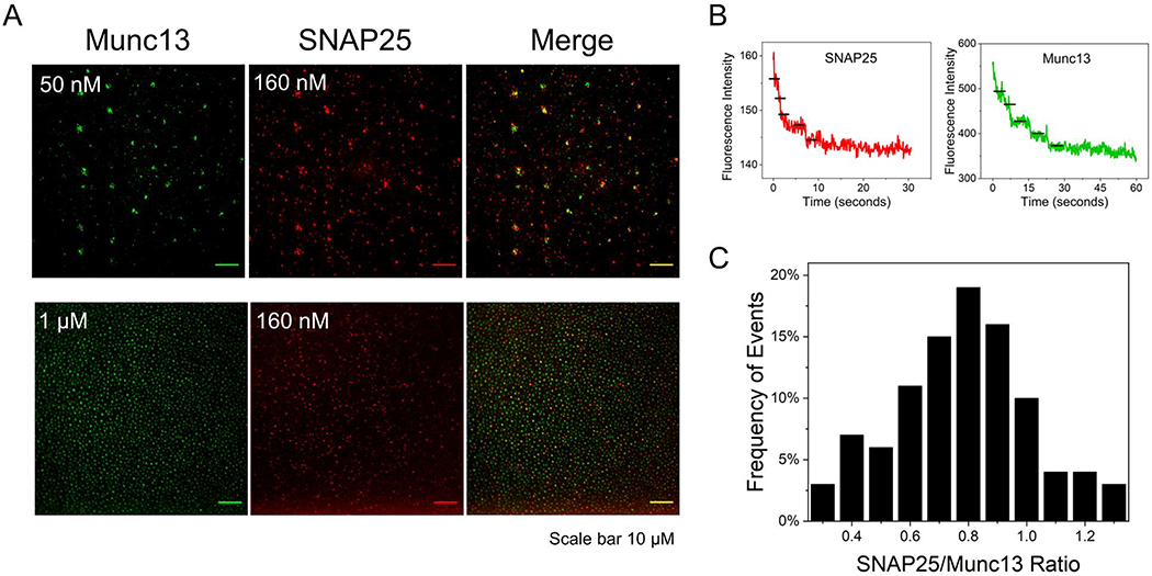
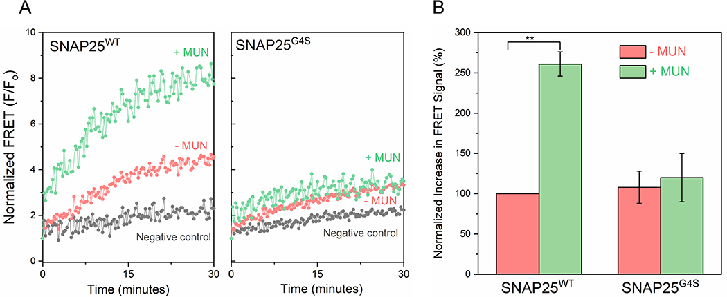
Synaptic vesicle fusion is mediated by SNARE proteins-VAMP2 on the vesicle and Syntaxin-1/SNAP25 on the presynaptic membrane. Chaperones Munc18-1 and Munc13-1 cooperatively catalyze SNARE assembly via an intermediate 'template' complex containing Syntaxin-1 and VAMP2. How SNAP25 enters this reaction remains a mystery. Here, we report that Munc13-1 recruits SNAP25 to initiate the ternary SNARE complex assembly by direct binding, as judged by bulk FRET spectroscopy and single-molecule optical tweezer studies. Detailed structure-function analyses show that the binding is mediated by the Munc13-1 MUN domain and is specific for the SNAP25 'linker' region that connects the two SNARE motifs. Consequently, freely diffusing SNAP25 molecules on phospholipid bilayers are concentrated and bound in ~ 1 : 1 stoichiometry by the self-assembled Munc13-1 nanoclusters.
Figure 1.. Dissecting Munc13–1 interaction with SNAP25…
Figure 1.. Dissecting Munc13–1 interaction with SNAP25 using Microscale Thermophoresis (MST) analysis.
(A) Titration of…
Figure 1.. Dissecting Munc13–1 interaction with SNAP25 using Microscale Thermophoresis (MST) analysis.
(A) Titration of full-length SNAP25 into AlexaFluor 660 labeled Munc13L (C1-C2B-MUN-C2C domains) produced a concentration-dependent change in the MST signal, yielding an apparent dissociation constant (Kd) = 26 ± 2 μM implying that Munc13L directly binds to SNAP25. Munc13–1 domain likely binds to the SNAP25 linker region as replacing the SNAP25 linker domain that connects that SNARE helical motifs with a GGGGS sequence (SNAP25G4S) completely abrogates Munc13-SNAP25 interaction (red curve). This interaction is highly specific as swapping the SNAP25 linker region with closely associated SNAP23 linker (SNAP25SNAP23) also disrupts the binding (blue curve).(B) MUN domain (orange curve) alone is sufficient to bind SNAP25 albeit with slightly reduced affinity (Kd = 48 ± 3 μM) and SNAP25 binding site is unique and different from the Syntaxin-1 binding site, as the Syntaxin non-binding Munc13–1 mutant (Munc13LNFAA) is capable of binding SNAP25 (purple curve). Average and standard deviations from a minimum of three independent experiments are shown.
Figure 2.. Munc13–1 binds and clusters SNAP25…
Figure 2.. Munc13–1 binds and clusters SNAP25 on lipid bilayer surface.
(A) Distribution of AlexaFluor…
Figure 2.. Munc13–1 binds and clusters SNAP25 on lipid bilayer surface.
(A) Distribution of AlexaFluor 488 labeled SNAP25WT (~150 nM) on supported lipid bilayer under physiological lipid and buffer composition in the absence or presence of Munc13L was visualized using TIRF microscopy. Munc13L induces clustering of SNAP25WT molecules and the number/size of the SNAP25 clusters directly correlates to concentration of Munc13L added. (B) Munc13L fails to cluster SNAP25 linker domain mutant (SNAP25G4S) even at the highest concentration (2 μM) tested. This suggests that MUN-SNAP25 linker domain interaction is feasible on lipid bilayers surfaces and results in clustering of otherwise diffusely distributed SNAP25 molecules. Representative fluorescent images are shown.
Figure 3.. Munc13–1 co-clusters with SNAP25 at…
Figure 3.. Munc13–1 co-clusters with SNAP25 at 1:1 stoichiometry.
(A) Representative fluorescence images showing that AlexaFluor 555 labeled SNAP25WT co-localizes with clusters of AlexaFluor 488 labeled Munc13L on lipid bilayer surface and the extent of co-localization depends on the concentration of Munc13L added. SNAP25 (160 nM) shows limited co-localization (Manders’ coefficient ~ 0.2) at 50 nM of Munc13L but has high overlap (Manders’ coefficient ~ 0.7) at 1μM of Munc13L. (B) Stoichiometry of SNAP25 and Munc13L in the co-clusters under low Munc13L concentration was estimated using step-bleaching analysis. Representative step-bleaching traces for SNAP25 (red) and Munc13L (green) are shown and the ratio of SNAP25:Munc13L quantified from ~70 co-clusters is shown as the histogram. Under these conditions, each cluster contains ~5–7 molecules at almost 1:1 ratio of Munc13L:SNAP25. This indicates that molecular interaction between the MUN domain and the SNAP25 linker region is likely responsible for the observed co-clustering of Munc13–1 and SNAP25.
Figure 4.. Munc13 interaction with SNAP25 is…
Figure 4.. Munc13 interaction with SNAP25 is essential for its chaperone function.
FRET between AlexaFluor…
Figure 4.. Munc13 interaction with SNAP25 is essential for its chaperone function.
FRET between AlexaFluor 488 labeled SNAP25 and AlexaFluor 555 labeled soluble VAMP2 introduced in the N-terminus (SNAP25 residue 20 & VAMP2 residue 28) shows that in the presence of cytoplasmic Syntaxin-1, the MUN domain activates the initial engagement of the SNARE complex. Negative control wherein Syntaxin-1 was left out confirms the observed FRET signal corresponds to formation of the ternary SNARE complex. The stimulatory effect of MUN domain was not observed with SNAP25 linker mutant (SNAP25G4S) indicating that the MUN-SNAP25 linker domain interaction is crucial for Munc13–1 ability to promote SNARE complex formation. Representative fluorescence traces for first 30 min of the reaction (left) and end-point quantification at 60 min (right) from two independent trials are shown. Statistical significance was determined using student t-test (** p<0.005).
Figure 5:. Munc13–1 recruits SNAP25 to Munc18–1/Syntaxin-1/VAMP2…
Figure 5:. Munc13–1 recruits SNAP25 to Munc18–1/Syntaxin-1/VAMP2 template complex.
(A) Experimental setup for optical tweezers.…
Figure 5:. Munc13–1 recruits SNAP25 to Munc18–1/Syntaxin-1/VAMP2 template complex.
(A) Experimental setup for optical tweezers. A single SNARE complex was pulled from the C termini of Syntaxin 1A (red) and VAMP2 (blue) via two DNA handles attached to two optically trapped polystyrene beads. The N-termini of Syntaxin 1A and VAMP2 were cross-linked via a disulfide bond. Munc18–1 (gray) and the MUN domain of Munc13–1 (yellow) were added in the solution. (B) Representative force extension curves (FECs) obtained for SNARE complexes assembled with SNAP25WT or SNAP25G4S in the presence of Munc18–1 and MUN domain as indicated. The Syntaxin–VAMPs conjugate was pulled (grey FECs) or relaxed (black FECs) by changing the separation between two optical traps at a speed of 10 nm/s or held at constant mean force around 5 pN (red FECs). The molecular states associated with different FEC regions are indicated by the corresponding state numbers (see Supplementary Figure 5 for their molecular diagrams) (C) Probability of full SNARE complex assembly from the Munc18–1/Syntaxin-1/VAMP2 template complex observed within 100s at 5 pN constant mean force. MUN domain ability to stimulate SNARE complex assembly is significantly diminished with the SNAP25 linker domain mutant (SNAP25G4S) as compared to SNAP25WT (p = 0.065). The N value refers to the total number of trials or pulling/relaxation rounds and average and standard error of the means are shown the error bar indicates the standard deviation (σ) of the corresponding probability (p) calculated as σ = √(p(1−p)/N).
Jahn R and Fasshauer D, Molecular machines governing exocytosis of synaptic vesicles. Nature, 2012. 490(7419): p. 201–7.
-
PMC
-
PubMed
Rizo J and Xu J, The Synaptic Vesicle Release Machinery. Annu Rev Biophys, 2015. 44: p. 339–67.
-
PubMed
Sudhof TC, Neurotransmitter release: the last millisecond in the life of a synaptic vesicle. Neuron, 2013. 80(3): p. 675–90.
-
PMC
-
PubMed
Sudhof TC and Rothman JE, Membrane fusion: grappling with SNARE and SM proteins. Science, 2009. 323(5913): p. 474–7.
-
PMC
-
PubMed
Sollner T, et al., A protein assembly-disassembly pathway in vitro that may correspond to sequential steps of synaptic vesicle docking, activation, and fusion. Cell, 1993. 75(3): p. 409–18.
-
PubMed