. 2021 Jan;70(1):37-48.
doi: 10.2144/btn-2020-0119. Epub 2020 Nov 23.

Generation of genetically tailored porcine liver cancer cells by CRISPR/Cas9 editing

Affiliations

Generation of genetically tailored porcine liver cancer cells by CRISPR/Cas9 editing

Lobna Elkhadragy et al. Biotechniques. 2021 Jan.

Abstract

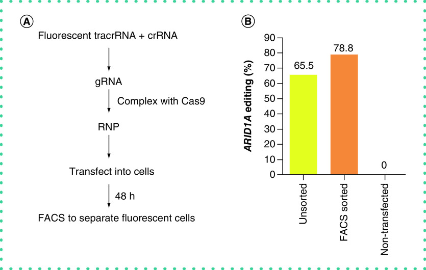
Pigs provide a valuable large animal model for several diseases due to their similarity with humans in anatomy, physiology, genetics and drug metabolism. We recently generated a porcine model for TP53R167H and KRASG12D driven hepatocellular carcinoma (HCC) by autologous liver implantation. Here we describe a streamlined approach for developing genetically tailored porcine HCC cells by CRISPR/Cas9 gene editing and isolation of homogenous genetically validated cell clones. The combination of CRISPR/Cas9 editing of HCC cells described herein with the orthotopic HCC model enables development of various porcine HCC models, each with a specific mutational profile. This allows modeling the effect of different driver mutation combinations on tumor progression and in vivo testing of novel targeted therapeutic approaches in a clinically relevant large animal model.

Keywords: CRISPR/Cas9; gene editing; gene knockout; hepatocellular carcinoma; large animal model; liver cancer; porcine cells.


Conflict of interest statement

Financial & competing interests disclosure

This work was supported by the National Institutes of Health – National Cancer Institute (1R21CA219461-01A1), US Department of Defense (Translational Team Science Award CA150590) and the Department of Radiology, University of Illinois at Chicago. L Schook, R Gaba and K Schachtschneider have received research support from Guerbet USA LLC, Janssen Research & Development LLC, the US Department of Defense and the US National Institutes of Health, and are scientific consultants for Sus Clinicals, Inc. L Elkhadragy, M Regan, W Totura, K Dasteh Goli, S Patel, K Garcia and M Stewart do not have conflicts of interest. The authors have no other relevant affiliations or financial involvement with any organization or entity with a financial interest in or financial conflict with the subject matter or materials discussed in the manuscript apart from those disclosed.

No writing assistance was utilized in the production of this manuscript.

Figures

Figure 1.
Figure 1.. Schematic representation of the workflow to generate porcine gene knockout cells using CRISPR/Cas9.
A ribonucleoprotein (RNP) complex is formed by incubating gRNA with Cas9. The RNP complex is transfected into cells using lipid nanoparticles resulting in a diverse array of mutations at the genomic target site in the cells. By isolation and sequence analysis of single cell clones, clonally homogenous cell lines with the desired edits can be obtained. NGS: Next-generation sequencing.
Figure 2.
Figure 2.. Isolation of hepatocytes from porcine liver section.
(A & B) A section of porcine liver is exposed and resected. (C) Cannulation of visible blood vessels on the cut surface of the liver section. (D) The liver section becomes pale after perfusion. Nonperfused red regions of the liver are discarded.
Figure 3.
Figure 3.. CRISPR/Cas9-mediated disruption of the porcine ARID1A gene.
(A) Schematic representation of the porcine ARID1A locus, showing the location of spacer sequences crRNA#1 and crRNA#2 (underlined blue font). Protospacer-adjacent motif sequences are marked in red. The introns are represented by lines, coding regions of exons are shaded in black and the untranslated region is white. (B) Comparing the CRISPR/Cas9-mediated editing efficiency of two ARID1A-targeting gRNAs. Porcine HCC line-1 cells were transfected with 15 nM RNP comprising Cas9 and gRNA#1 or gRNA#2. Nontransfected cells were used as control. Genomic DNA was collected 2 days post-transfection and analyzed by targeted NGS. The bar graph depicts the percentages (%) of total reads that displayed indels at the gRNA target site occurring as a result of nonhomologous end joining. (C & D) Top 15 reads detected by targeted NGS analysis, mapped to the reference sequence. The percentages of reads for each sequence are shown on the right. The asterisk indicates non-edited reads. Dashed line: predicted Cas9 cleavage position; red box: insertion; dash: deleted base. HCC: Hepatocellular carcinoma; NGS: Next-generation sequencing; RNP: Ribonucleoprotein.
Figure 4.
Figure 4.. Fluorescence-activated cell sorting increases the percentage of gene-edited cells.
(A) Schematic representation of the workflow to sort RNP-transfected cells. A tracrRNA conjugated with ATTO-550 fluorescent dye was mixed with crRNA to form gRNA. The fluorescently labeled gRNA and purified Cas9 were complexed to form a ribonucleoprotein complex (RNP) which was then transfected into cells, allowing visual assessment of transfection efficiency using a fluorescence microscope. 48 h later, the cells were sorted by FACS to separate successfully transfected fluorescent cells. (B) ARID1A editing efficiency in unsorted or FACS-sorted porcine HCC cells. Porcine HCC line-1 cells were transfected with 15 nM RNP comprising Cas9 and ARID1A targeting gRNA#1. Nontransfected control cells were also analyzed. FACS-sorted cells were used for genomic analysis directly after sorting. The bar graph depicts the percentages (%) of total reads that displayed indels at the gRNA target site as analyzed by NGS. HCC: Hepatocellular carcinoma; NGS: Next-generation sequencing.
Figure 5.
Figure 5.. Increased CRISPR-Cas9 editing efficiency of ARID1A at higher ribonucleoprotein concentration.
(A) ARID1A editing efficiency in four porcine HCC cell lines following transfection with 25 nM ribonucleoprotein comprising Cas9 and gRNA#1. Genomic DNA was collected 2 days post-transfection and analyzed by targeted NGS. The bar graph depicts the percentages (%) of total reads that displayed indels at the gRNA target site occurring as a result of nonhomologous end joining. (B) Top 15 reads detected by targeted NGS analysis in the four porcine HCC lines, mapped to the reference sequence. The identity and frequency of indels in the four cell lines is consistent. The percentages of reads of each sequence are shown on the right. The location of the indel relative to the expected cleavage site is shown on the left, followed by a colon and the number of nucleotides inserted (i) or deleted (d). The asterisk denotes non-edited reads. Dashed line: predicted Cas9 cleavage position; red box: insertion; dash: deleted base. HCC: Hepatocellular carcinoma; NGS: Next-generation sequencing.
Figure 6.
Figure 6.. Comparing analysis of CRISPR/Cas9 mediated disruption of ARID1A by next-generation and Sanger sequencing.
(A) Sanger sequencing chromatograms of porcine HCC cells transfected with Cas9 and ARID1A gRNA#1 and control nontransfected cells. Overlapping peaks near the gRNA target site are observed for the sample subjected to CRISPR/Cas9 editing. Dashed line: predicted Cas9 cleavage position; underlined nucleotides: crRNA spacer sequence. (B) Top 5 reads detected by ICE software analysis of Sanger sequencing data in three porcine HCC lines transfected with Cas9 and ARID1A gRNA#1. The type and frequency of indels is shown on the left and is consistent among the three cell lines. (C) The editing efficiency (%) and frequency of the top five frequent reads (%) identified by ICE software analysis of Sanger sequencing data as compared with NGS analysis in three ARID1A-edited porcine HCC cells. The location of the indel relative to the expected cleavage site is shown on the left, followed by a colon and the number of nucleotides inserted (i) or deleted (d). Overall similarity was noted between the two analysis methods. HCC: Hepatocellular carcinoma; NGS: Next-generation sequencing.
Figure 7.
Figure 7.. Representative single cell clones isolated from porcine hepatocellular carcinoma cells subject to CRISPR/Cas9-mediated ARID1A disruption.
(A) Possible editing outcomes of CRISPR/Cas9 include the following: an identical indel is introduced in both alleles (homozygous mutation), different indels occur in each allele (biallelic mutation) or an indel is observed in an allele while the other allele remains unedited (heterozygous mutation). (B–D) Analysis of representative single cells clones isolated from porcine HCC cells transfected with Cas9 and ARID1A gRNA#1. (B) Reads detected by targeted NGS analysis mapped to the reference sequence (top) for three single cell clones. Dashed line: predicted Cas9 cleavage position; red box: insertion; dash: deleted base. (C) The predicted translation of ARID1A protein for the representative clones. The dotted region represents amino acids with frameshift mutation. (D) Arginase-1 staining (green) merged with DAPI staining (blue) for parental porcine HCC cell line and the representative single-cell clones (scale bar = 200 μm). All clones show positive arginase-1 staining, confirming their hepatocellular origin. HCC: Hepatocellular carcinoma; NGS: Next-generation sequencing.

References

    1. Flisikowska T, Kind A, Schnieke A Pigs as models of human cancers. Theriogenology 86(1), 433–437 (2016). - PubMed
    1. Gun G, Kues WA Current progress of genetically engineered pig models for biomedical research. Biores. Open Access 3(6), 255–264 (2014). - PMC - PubMed
    1. Hryhorowicz M, Lipinski D, Hryhorowicz S, Nowak-Terpilowska A, Ryczek N, Zeyland J Application of genetically engineered pigs in biomedical research. Genes (Basel) 11(6), 670 (2020). - PMC - PubMed
    1. Schachtschneider KM, Schwind RM, Darfour-Oduro KA et al. A validated, transitional and translational porcine model of hepatocellular carcinoma. Oncotarget 8(38), 63620–63634 (2017). - PMC - PubMed
    1. Gaba RC, Elkhadragy L, Boas FE et al. Development and comprehensive characterization of porcine hepatocellular carcinoma for translational liver cancer investigation. Oncotarget 11, 2686–2701 (2020). - PMC - PubMed

Publication types