. 2020 Nov 23;15(1):68.
doi: 10.1186/s13024-020-00414-3.

Regional binding of tau and amyloid PET tracers in Down syndrome autopsy brain tissue

Affiliations

Regional binding of tau and amyloid PET tracers in Down syndrome autopsy brain tissue

L Lemoine et al. Mol Neurodegener. .

Abstract

Introduction: Tau pathology is a major age-related event in Down syndrome with Alzheimer's disease (DS-AD). Although recently, several different Tau PET tracers have been developed as biomarkers for AD, these tracers showed different binding properties in Alzheimer disease and other non-AD tauopathies. They have not been yet investigated in tissue obtained postmortem for DS-AD cases. Here, we evaluated the binding characteristics of two Tau PET tracers (3H-MK6240 and 3H-THK5117) and one amyloid (3H-PIB) ligand in the medial frontal gyrus (MFG) and hippocampus (HIPP) in tissue from adults with DS-AD and DS cases with mild cognitive impairment (MCI) compared to sporadic AD.

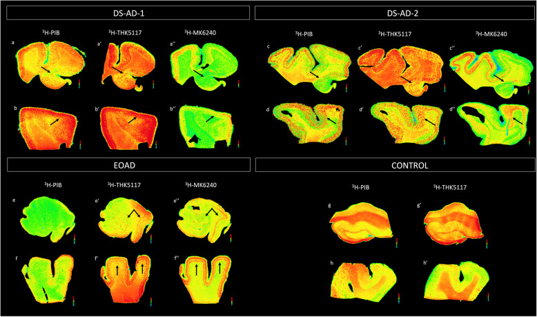
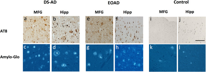
Methods: Tau and amyloid autoradiography were performed on paraffin-embedded sections. To confirm respective ligand targets, adjacent sections were immunoreacted for phospho-Tau (AT8) and stained for amyloid staining using Amylo-Glo.

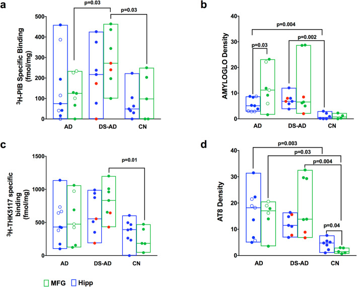
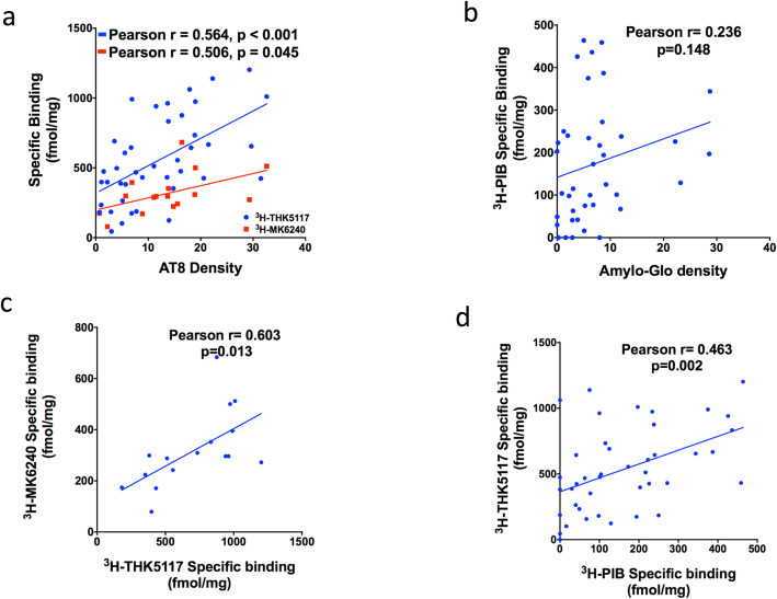
Results: The two Tau tracers showed a significant correlation with each other and with AT8, suggesting that both tracers were binding to Tau deposits. 3H-MK6240 Tau binding correlated with AT8 immunostaining but to a lesser degree than the 3H-THK5117 tracer, suggesting differences in binding sites between the two Tau tracers. 3H-THK5117, 3H-MK6240 and 3H-PIB displayed dense laminar binding in the HIPP and MFG in adult DS brains. A regional difference in Tau binding between adult DS and AD was observed suggesting differential regional Tau deposition in adult DS compared to AD, with higher THK binding density in the MFG in adult with DS compared to AD. No significant correlation was found between 3H-PIB and Amylo-Glo staining in adult DS brains suggesting that the amyloid PIB tracer binds to additional sites.

Conclusions: This study provides new insights into the regional binding distribution of a first-generation and a second-generation Tau tracer in limbic and neocortical regions in adults with DS, as well as regional differences in Tau binding in adult with DS vs. those with AD. These findings provide new information about the binding properties of two Tau radiotracers for the detection of Tau pathology in adults with DS in vivo and provide valuable data regarding Tau vs. amyloid binding in adult DS compared to AD.

Keywords: Alzheimer’s disease; Down syndrome; Neurofibrillary tangles; Neuropathology; PET tracers.


Conflict of interest statement

The authors do not report any conflicts of interest.

Figures

Fig. 1
Fig. 1
Amyloid and Tau autoradiography binding in 2 DS-AD in comparison to 1 EOAD and 1 control. Top row: DS-AD-1 57-year-old female with DS-AD. From left to right: Autoradiographic images showing 3H-PIB binding (a, b),3H-THK5117 binding (a’,b’) and 3H-MK6240 binding (a”,b”) on an adjacent section in HIPP and MFG. DS-AD-2 62-year-old female with DS-AD From left to right: Autoradiographic images showing 3H-PIB binding (c,d),3H-THK5117 binding (c’,d’) and 3H-MK6240 binding (c”,d”) on an adjacent section in HIPP and MFG. Bottom row: EOAD: 55-year-old female diagnosed with Alzheimer’s disease: From left to right: Autoradiographic images showing 3H-PIB binding (e,f),3H-THK5117 binding (e’,f’) and 3H-MK6240 binding (e”,f”) on an adjacent section in HIPP and MFG. CONTROL: 59-year-old control case. From left to right: Autoradiographic images showing 3H-PIB binding (g, h),3H-THK5117 binding (g’,h’) on an adjacent section in HIPP and MFG
Fig. 2
Fig. 2
Amylo-Glo and AT8 immunostaining in HIPP and MFG of 1 DS-AD, 1 EOAD and 1 control case. From left to right: AT8 staining in DS-AD (a,b) and Amylo-Glo staining (c,d) in MFG and HIPP respectively. AT8 staining in 1 EOAD (e,f) and Amylo-Glo staining (g,h) in MFG and HIPP respectively. AT8 staining in 1 control (i,j) and Amylo-Glo staining (k,l) in MFG and HIPP respectively
Fig. 3
Fig. 3
Semi-quantitative analysis of 3H-PiB, 3H-THK5117 autoradiography and Amylo-Glo and AT8 staining. Box plot with a line at the median represent: a: Specific binding of 3H-PIB and for AD, DS and CN in HIPP and MFG. b: Amylo-Glo density for AD, DS and CN in HIPP and MFG. c: Specific binding of 3H-THK5117 for AD, DS and CN in HIPP and MFG. d: AT8 density for AD, DS and CN in HIPP and MFG. Specific binding for 3H-PIB and 3H-THK5117 is represented in fmol/mg. Gray matter was delineated manually using multigauge software. Density values for Amylo-Glo and AT8 are presented in b and d respectively. The red dots in each plot represent two DS-MCI cases. The empty blue circles in AD represent the EOAD
Fig. 4
Fig. 4
Linear regressions between autoradiography binding and density staining. a. 3H-THK5117 and AT8 immunostaining (Pearson r = 0.564, p < 0.001, shown in blue), and 3H-MK6240 and AT8 immunostaining (Pearson r = 0.506, p = 0.045, shown in red) in the whole cohort (AD, DS-AD, CN) in both regions (MFG + HIPP). b: 3H-PIB and Amylo-Glo (Pearson r = 0.236, p = 0.148) in the whole population (AD, DS-AD,CN) in both regions (MFG + HIPP). c: 3H-THK5117 and 3H-MK6240 (Pearson r = 0.603, p = 0.013) in the whole population (AD, DS-AD,CN) in both regions (MFG + HIPP). d: Correlation in the whole population (AD,DS-AD,CN) in both regions (MFG + HIPP) between 3H-PIB and 3H-THK5117(Pearson r = 0.463, p = 0.002)

Similar articles

Cited by

References

    1. Hartley D, Blumenthal T, Carrillo M, DiPaolo G, Esralew L, Gardiner K, Granholm AC, Iqbal K, Krams M, Lemere C, et al. Down syndrome and Alzheimer's disease: common pathways, common goals. Alzheimers Dement. 2015;11:700–709. doi: 10.1016/j.jalz.2014.10.007. - DOI - PMC - PubMed
    1. Head E, Garzon-Rodriguez W, Johnson JK, Lott IT, Cotman CW, Glabe C. Oxidation of Abeta and plaque biogenesis in Alzheimer's disease and Down syndrome. Neurobiol Dis. 2001;8:792–806. doi: 10.1006/nbdi.2001.0431. - DOI - PubMed
    1. Head E, Lott IT, Wilcock DM, Lemere CA. Aging in Down syndrome and the development of Alzheimer's disease neuropathology. Curr Alzheimer Res. 2016;13:18–29. doi: 10.2174/1567205012666151020114607. - DOI - PMC - PubMed
    1. Prasher VP, Sajith SG, Mehta P, Zigman WB, Schupf N. Plasma beta-amyloid and duration of Alzheimer's disease in adults with Down syndrome. Int J Geriatr Psychiatry. 2010;25:202–207. doi: 10.1002/gps.2321. - DOI - PMC - PubMed
    1. Firth NC, Startin CM, Hithersay R, Hamburg S, Wijeratne PA, Mok KY, Hardy J, Alexander DC, LonDown SC, Strydom A. Aging related cognitive changes associated with Alzheimer's disease in Down syndrome. Ann Clin Transl Neurol. 2018;5:741–751. doi: 10.1002/acn3.571. - DOI - PMC - PubMed

Publication types