In vivo bioluminescence tomography-guided radiation research platform for pancreatic cancer: an initial study using subcutaneous and orthotopic pancreatic tumor models
- PMID: 33223595
- PMCID: PMC7677029
- DOI: 10.1117/12.2546503
In vivo bioluminescence tomography-guided radiation research platform for pancreatic cancer: an initial study using subcutaneous and orthotopic pancreatic tumor models
Abstract
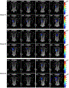
Genetically engineered mouse model(GEMM) that develops pancreatic ductal adenocarcinoma(PDAC) offers an experimental system to advance our understanding of radiotherapy(RT) for pancreatic cancer. Cone beam CT(CBCT)-guided small animal radiation research platform(SARRP) has been developed to mimic the RT used for human. However, we recognized that CBCT is inadequate to localize the PDAC growing in low image contrast environment. We innovated bioluminescence tomography(BLT) to guide SARRP irradiation for in vivo PDAC. Before working on the complex PDAC-GEMM, we first validated our BLT target localization using subcutaneous and orthotopic pancreatic tumor models. Our BLT process involves the animal transport between the BLT system and SARRP. We inserted a titanium wire into the orthotopic tumor as the fiducial marker to track the tumor location and to validate the BLT reconstruction accuracy. Our data shows that with careful animal handling, minimum disturbance for target position was introduced during our BLT imaging procedure(<0.5mm). However, from longitudinal 2D bioluminescence image(BLI) study, the day-to-day location variation for an abdominal tumor can be significant. We also showed that the 2D BLI in single projection setting cannot accurately capture the abdominal tumor location. It renders that 3D BLT with multiple-projection is needed to quantify the tumor volume and location for precise radiation research. Our initial results show the BLT can retrieve the location at 2mm accuracy for both tumor models, and the tumor volume can be delineated within 25% accuracy. The study for the subcutaneous and orthotopic models will provide us valuable knowledge for BLT-guided PDAC-GEMM radiation research.
Keywords: bioluminescence tomography; image-guided radiation therapy; pancreatic cancer; small animal irradiator.
Figures


Similar articles
-
Characterization of a commercial bioluminescence tomography-guided system for pre-clinical radiation research.Med Phys. 2023 Oct;50(10):6433-6453. doi: 10.1002/mp.16669. Epub 2023 Aug 26. Med Phys. 2023. PMID: 37633836 Free PMC article.
-
In vivo bioluminescence tomography-guided system for pancreatic cancer radiotherapy research.Biomed Opt Express. 2024 Jul 9;15(8):4525-4539. doi: 10.1364/BOE.523916. eCollection 2024 Aug 1. Biomed Opt Express. 2024. PMID: 39347008 Free PMC article.
-
In Vivo Bioluminescence Tomography Center of Mass-Guided Conformal Irradiation.Int J Radiat Oncol Biol Phys. 2020 Mar 1;106(3):612-620. doi: 10.1016/j.ijrobp.2019.11.003. Epub 2019 Nov 15. Int J Radiat Oncol Biol Phys. 2020. PMID: 31738948 Free PMC article.
-
Pancreatic stellate cells and pancreas cancer: current perspectives and future strategies.Eur J Cancer. 2014 Oct;50(15):2570-82. doi: 10.1016/j.ejca.2014.06.021. Epub 2014 Aug 1. Eur J Cancer. 2014. PMID: 25091797 Review.
-
Advances in 4D medical imaging and 4D radiation therapy.Technol Cancer Res Treat. 2008 Feb;7(1):67-81. doi: 10.1177/153303460800700109. Technol Cancer Res Treat. 2008. PMID: 18198927 Review.
Cited by
-
Mobile bioluminescence tomography-guided system for pre-clinical radiotherapy research.Biomed Opt Express. 2022 Aug 30;13(9):4970-4989. doi: 10.1364/BOE.460737. eCollection 2022 Sep 1. Biomed Opt Express. 2022. PMID: 36187243 Free PMC article.
-
Quantitative molecular bioluminescence tomography.J Biomed Opt. 2022 Jun;27(6):066004. doi: 10.1117/1.JBO.27.6.066004. J Biomed Opt. 2022. PMID: 35726130 Free PMC article.
-
Characterization of a commercial bioluminescence tomography-guided system for pre-clinical radiation research.Med Phys. 2023 Oct;50(10):6433-6453. doi: 10.1002/mp.16669. Epub 2023 Aug 26. Med Phys. 2023. PMID: 37633836 Free PMC article.
-
Histopathological Tumor and Normal Tissue Responses after 3D-Planned Arc Radiotherapy in an Orthotopic Xenograft Mouse Model of Human Pancreatic Cancer.Cancers (Basel). 2021 Nov 12;13(22):5656. doi: 10.3390/cancers13225656. Cancers (Basel). 2021. PMID: 34830813 Free PMC article.
References
-
- Bray F, Ferlay J, Soerjomataram I, Siegel RL, Torre LA, and Jemal A, “Global cancer statistics 2018: GLOBOCAN estimates of incidence and mortality worldwide for 36 cancers in 185 countries,” CA-Cancer J. Clin 68(6), 394–424 (2018). - PubMed
-
- “Chemoimmunotherapy and radiation in pancreatic cancer (CRIT),” ClinicalTrials.gov. Identifier: NCT01903083.
-
- “Pancreatic tumor cell vaccine (GVAX), low dose cyclophosphamide, fractionated stereotactic body radiation therapy (SBRT), and FOLFIRINOX chemotherapy in patients with resected adenocarcinoma of the pancreas,” ClinicalTrials.gov. Identifier: NCT01595321.
-
- “Immune checkpoint inhibition (Tremelimumab and/or MEDI4736) in combination with radiation therapy in patients with unresectable pancreatic cancer,” ClinicalTrials.gov. Identifier: NCT02311361.
-
- Wong J, Armour E, Kazanzides P, Iordachita U, Tryggestad E, Deng H, Matinfar M, Kennedy C, Liu ZJ, Chan T, Gray O, Verhaegen F, McNutt T, Ford E, and DeWeese TL, “High-resolution, small animal radiation research platform with X-ray tomographic guidance capabilities,” Int. J. Radiat. Oncol. Biol. Phys 71(5), 1591–1599 (2008). - PMC - PubMed
Grants and funding
LinkOut - more resources
Full Text Sources
