. 2020 Nov 2:13:522073.
doi: 10.3389/fnmol.2020.552073. eCollection 2020.

The Interplay Between Beta-Amyloid 1-42 (Aβ1-42)-Induced Hippocampal Inflammatory Response, p-tau, Vascular Pathology, and Their Synergistic Contributions to Neuronal Death and Behavioral Deficits

Affiliations

The Interplay Between Beta-Amyloid 1-42 (Aβ1-42)-Induced Hippocampal Inflammatory Response, p-tau, Vascular Pathology, and Their Synergistic Contributions to Neuronal Death and Behavioral Deficits

Beatriz Calvo-Flores Guzmán et al. Front Mol Neurosci. .

Abstract

Alzheimer's disease (AD), the most common chronic neurodegenerative disorder, has complex neuropathology. The principal neuropathological hallmarks of the disease are the deposition of extracellular β-amyloid (Aβ) plaques and neurofibrillary tangles (NFTs) comprised of hyperphosphorylated tau (p-tau) protein. These changes occur with neuroinflammation, a compromised blood-brain barrier (BBB) integrity, and neuronal synaptic dysfunction, all of which ultimately lead to neuronal cell loss and cognitive deficits in AD. Aβ1-42 was stereotaxically administered bilaterally into the CA1 region of the hippocampi of 18-month-old male C57BL/6 mice. This study aimed to characterize, utilizing immunohistochemistry and behavioral testing, the spatial and temporal effects of Aβ1-42 on a broad set of parameters characteristic of AD: p-tau, neuroinflammation, vascular pathology, pyramidal cell survival, and behavior. Three days after Aβ1-42 injection and before significant neuronal cell loss was detected, acute neuroinflammatory and vascular responses were observed. These responses included the up-regulation of glial fibrillary acidic protein (GFAP), cell adhesion molecule-1 (PECAM-1, also known as CD31), fibrinogen labeling, and an increased number of activated astrocytes and microglia in the CA1 region of the hippocampus. From day 7, there was significant pyramidal cell loss in the CA1 region of the hippocampus, and by 30 days, significant localized up-regulation of p-tau, GFAP, Iba-1, CD31, and alpha-smooth muscle actin (α-SMA) in the Aβ1-42-injected mice compared with controls. These molecular changes in Aβ1-42-injected mice were accompanied by cognitive deterioration, as demonstrated by long-term spatial memory impairment. This study is reporting a comprehensive examination of a complex set of parameters associated with intrahippocampal administration of Aβ1-42 in mice, their spatiotemporal interactions and combined contribution to the disease progression. We show that a single Aβ injection can reproduce aspects of the inflammatory, vascular, and p-tau induced pathology occurring in the AD human brain that lead to cognitive deficits.

Keywords: Alzheiemer’s disease; cognition; neuroinflamamation; tau phosphorylation; β-amyloid.


Figures

Figure 1
Figure 1
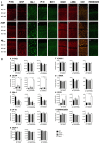
Panel I: Western blot against mouse brain protein homogenates probed with p-tau (A), glial fibrillary acidic protein (GFAP; B), Iba-1 (C), and ICAM-1 (D). Observed band sizes: p-tau ~58 kDA, GFAP ~50 kDA, Iba-1 ~17 kDA and ICAM-1 ~85 kDA. Panel II: Representative images showing immunolabeling of p-tau, GFAP, Iba-1, CD31, and fibrinogen at the injection site, 3 days post-injection in NC (a–e), ACSF- (f–j), and Aβ1–42-injected (k–o) mice. Scale bar (40 μm). Panel III: Graphs showing quantification at the injection site in the CA1 hippocampal region of p-tau, GFAP, Iba-1, CD31, and fibrinogen immunolabeling density, 3 days post-injection in NC, ACSF-, scrAβ1–42- and Aβ1–42-injected mice. Data expressed as mean ± SEM (Kruskal–Wallis test; *p < 0.05, n = 3–5).
Figure 2
Figure 2
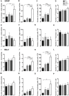
Graphs showing quantification of astrocytic and microglial morphology at the injection site (A–D,I–L) and a location adjacent to the injection site (E–H,M–P) in the CA1 hippocampal region 3 days post-injection in NC, ACSF-, scrAβ1–42- and Aβ1–42-injected mice. Data expressed as mean ± SEM (Kruskal–Wallis test; *p < 0.05; **p < 0.01, ***p < 0.001, ****p < 0.0001, n = 3–6).
Figure 3
Figure 3
Panel I: Representative images showing immunolabeling of p-tau, GFAP, Iba-1, IP-10, MCP-1, ICAM-1, α-SMA, CD31, and fibrinogen at a location adjacent to the injection site, 3 days after injection in NC (a–i), ACSF- (j–r) and Aβ1–42-injected (s–aa) mice. Scale bar (40 μm). Panel II: Graphs showing quantification at a location adjacent to the injection site in the CA1 hippocampal region of p-tau, GFAP, Iba-1, IP-10, MCP-1, ICAM-1, α-SMA, CD31, and fibrinogen (A–I) immunolabeling density, 3 days post-injection in NC, ACSF-, scrAβ1–42- and Aβ1–42-injected mice. Data expressed as mean ± SEM (Kruskal–Wallis test; *p < 0.05; **p < 0.01, ****p < 0.0001, n = 6).
Figure 4
Figure 4
Correlation between the number of activated astrocytes and microglia and vascular markers at the injection site (A–F, n = 3) and a location adjacent to the injection site (G–I, n = 6) in the CA1 hippocampal region 3 days post-injection in NC, ACSF-, scrAβ1–42- and Aβ1–42-injected mice. This relationship is represented by a Spearman’s r-value (*p < 0.05, **p < 0.01).
Figure 5
Figure 5
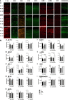
Panel I: Graphs showing the number of NeuN-positive pyramidal cells in the str. pyramidale of the CA1 hippocampal layer in NC, ACSF-, scrAβ1–42- and Aβ1–42-injected mice 7 days post-injection (A) and 30 days post-injection (B). Representative images of the CA1 hippocampal subregion at a location adjacent to the injection site showing NeuN-positive pyramidal cells from the str. pyramidale in NC, ACSF-, scrAβ1–42- and Aβ1–42-injected mice 30 days post-injection (C). Panel II: Representative images showing immunolabeling of p-tau, GFAP, Iba-1, CD31, and fibrinogen at the injection site 30 days post-injection in NC (a–e), ACSF-injected (f–j), and Aβ1–42-injected (k–o) mice. Scale bar (40 μm). Panel III: Graphs showing quantification at the injection site in the CA1 hippocampal region of p-tau, GFAP, Iba-1, CD31, and fibrinogen, 30 days post-injection in NC, ACSF-, scrAβ1–42- and Aβ1–42-injected mice. Data expressed as mean ± SEM (Kruskal–Wallis test; *p < 0.05; **p < 0.01, ****p < 0.0001, n = 6).
Figure 6
Figure 6
Graphs showing quantification of astrocytic and microglial morphology at the injection site (A–D,I–L) and a location adjacent to the injection site (E–H,M–P) in the CA1 hippocampal region 30 days post-injection in NC, ACSF-, scrAβ1–42- and Aβ1–42-injected mice. Data expressed as mean ± SEM (Kruskal–Wallis test; *p < 0.05, **p < 0.01, ***p < 0.001, ****p < 0.0001, n = 6).
Figure 7
Figure 7
Panel I: Representative images showing immunolabeling of p-tau, GFAP, Iba-1, IP-10, MCP-1, ICAM-1, a-SMA, CD31 and fibrinogen at a location adjacent to the injection site, 30 days post- injection in NC (a–i), ACSF-injected (j–r) and Aβ1–42-injected (s-aa) mice. Panel II: Graphs showing quantification at a location adjacent to the injection site in the CA1 hippocampal region of p-tau, GFAP, Iba-1, IP-10, MCP-1, ICAM-1, α-SMA, CD31 and fibrinogen immunolabeling density, 30 days post injection in NC, ACSF-, scrβ1–42- and Aβ1–42-injected mice. Data expressed as mean SEM (Kruskall-Wallis test; *p < 0.05; ***p < 0.001, n = 6).
Figure 8
Figure 8
Correlation between the number of activated astrocytes and microglia, p-tau, and vascular markers at the injection site (A–F) and at a location adjacent to the injection site (G–I) in the CA1 hippocampal region 30 days post-injection in NC, ACSF-, scrAβ1–42- and Aβ1–42-injected mice. This relationship is represented by a Spearman’s r-value (*p < 0.05, **p < 0.01, n = 6).
Figure 9
Figure 9
1–42-injected mice showed long-term spatial memory impairment revealed by the novel object alteration (A, performed on days 7, 8), novel object recognition (B, performed on days 11, 12), and Morris Water Maze (E, performed on days 20–24) tests. Short-term spatial memory assessed by the Y maze showed no significant difference in Aβ1–42-injected mice compared with controls (C, performed on day 15). Non-spatial memory remained unchanged in Aβ1–42-injected mice compared with controls (F, performed on day 27), and Aβ1–42 and/or the injection itself did not produce any anxiogenic effect (D, performed on day 17). Scatter plot showing all of the data values used with the median (Kruskal-Wallis test; *p < 0.05; **p < 0.01, ***p < 0.001, n = 12).
Figure 10
Figure 10
Correlation between the number of activated astrocytes and microglia (A–F), p-tau (G–I), CD31 (J–L)at the injection site in the CA1 hippocampal region, and behavioral scores 30 days post-injection in NC, ACSF-, scrAβ1–42- and Aβ1–42-injected mice. This relationship is represented by a Spearman’s r-value (*p < 0.05, n = 6).

Similar articles

Cited by

References

    1. Amaral D. G., Scharfman H. E., Lavenex P. (2007). The dentate gyrus: fundamental neuroanatomical organization (dentate gyrus for dummies). Prog. Brain Res. 163, 3–22. 10.1016/S0079-6123(07)63001-5 - DOI - PMC - PubMed
    1. Arbel-Ornath M., Hudry E., Boivin J. R., Hashimoto T., Takeda S., Kuchibhotla K. V., et al. (2017). Soluble oligomeric amyloid-β induces calcium dyshomeostasis that precedes synapse loss in the living mouse brain. Mol. Neurodegener. 12:27 10.1186/s13024-017-0169-928327181 - DOI - PMC - PubMed
    1. Arriagada P. V., Growdon J. H., Hedley-Whyte E. T., Hyman B. T. (1992). Neurofibrillary tangles but not senile plaques parallel duration and severity of Alzheimer’s disease. Neurology 42, 631–639. 10.1212/wnl.42.3.631 - DOI - PubMed
    1. Balducci C., Beeg M., Stravalaci M., Bastone A., Sclip A., Biasini E., et al. . (2010). Synthetic amyloid-β oligomers impair long-term memory independently of cellular prion protein. Proc. Natl. Acad. Sci. U S A 107, 2295–2300. 10.1073/pnas.0911829107 - DOI - PMC - PubMed
    1. Baluchnejadmojara D. T., Mohamadi-Zarch S., Roghani M. (2019). Safranal, an active ingredient of saffron, attenuates cognitive deficits in amyloid β-induced rat model of Alzheimer’s disease: underlying mechanisms metabolic brain disease. Metab. Brain Dis. 34, 1747–1759. 10.1073/pnas.0911829107 - DOI - PubMed