

Autophagy and Redox Homeostasis in Parkinson's: A Crucial Balancing Act
- PMID: 33224433
- PMCID: PMC7671810
- DOI: 10.1155/2020/8865611
Autophagy and Redox Homeostasis in Parkinson's: A Crucial Balancing Act
Abstract
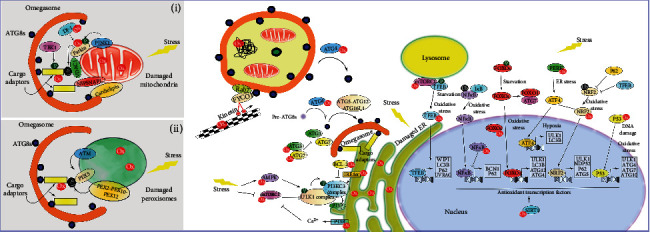
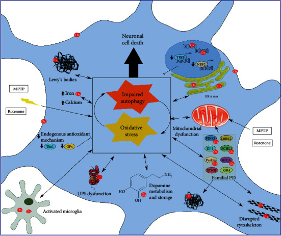
Reactive oxygen species (ROS) and reactive nitrogen species (RNS) are generated primarily from endogenous biochemical reactions in mitochondria, endoplasmic reticulum (ER), and peroxisomes. Typically, ROS/RNS correlate with oxidative damage and cell death; however, free radicals are also crucial for normal cellular functions, including supporting neuronal homeostasis. ROS/RNS levels influence and are influenced by antioxidant systems, including the catabolic autophagy pathways. Autophagy is an intracellular lysosomal degradation process by which invasive, damaged, or redundant cytoplasmic components, including microorganisms and defunct organelles, are removed to maintain cellular homeostasis. This process is particularly important in neurons that are required to cope with prolonged and sustained operational stress. Consequently, autophagy is a primary line of protection against neurodegenerative diseases. Parkinson's is caused by the loss of midbrain dopaminergic neurons (mDANs), resulting in progressive disruption of the nigrostriatal pathway, leading to motor, behavioural, and cognitive impairments. Mitochondrial dysfunction, with associated increases in oxidative stress, and declining proteostasis control, are key contributors during mDAN demise in Parkinson's. In this review, we analyse the crosstalk between autophagy and redoxtasis, including the molecular mechanisms involved and the detrimental effect of an imbalance in the pathogenesis of Parkinson's.
Copyright © 2020 Natalia Jimenez-Moreno and Jon D. Lane.
Conflict of interest statement
The authors declare that there is no conflict of interest regarding the publication of this paper.
Figures
Similar articles
-
Damage to dopaminergic neurons by oxidative stress in Parkinson's disease (Review).Int J Mol Med. 2018 Apr;41(4):1817-1825. doi: 10.3892/ijmm.2018.3406. Epub 2018 Jan 19. Int J Mol Med. 2018. PMID: 29393357 Review.
-
Mitochondria-mediated damage to dopaminergic neurons in Parkinson's disease (Review).Int J Mol Med. 2018 Feb;41(2):615-623. doi: 10.3892/ijmm.2017.3255. Epub 2017 Nov 16. Int J Mol Med. 2018. PMID: 29207041 Review.
-
Cholesterol contributes to dopamine-neuronal loss in MPTP mouse model of Parkinson's disease: Involvement of mitochondrial dysfunctions and oxidative stress.PLoS One. 2017 Feb 7;12(2):e0171285. doi: 10.1371/journal.pone.0171285. eCollection 2017. PLoS One. 2017. PMID: 28170429 Free PMC article.
-
Geraniol Protects Against the Protein and Oxidative Stress Induced by Rotenone in an In Vitro Model of Parkinson's Disease.Neurochem Res. 2018 Oct;43(10):1947-1962. doi: 10.1007/s11064-018-2617-5. Epub 2018 Aug 23. Neurochem Res. 2018. PMID: 30141137
-
Oxidative stress and autophagy in cardiovascular homeostasis.Antioxid Redox Signal. 2014 Jan 20;20(3):507-18. doi: 10.1089/ars.2013.5359. Epub 2013 Jun 27. Antioxid Redox Signal. 2014. PMID: 23641894 Free PMC article. Review.
Cited by
-
Rasagiline Exerts Neuroprotection towards Oxygen-Glucose-Deprivation/Reoxygenation-Induced GAPDH-Mediated Cell Death by Activating Akt/Nrf2 Signaling.Biomedicines. 2024 Jul 17;12(7):1592. doi: 10.3390/biomedicines12071592. Biomedicines. 2024. PMID: 39062165 Free PMC article.
-
Ataxia Telangiectasia Mutated Protein Kinase: A Potential Master Puppeteer of Oxidative Stress-Induced Metabolic Recycling.Oxid Med Cell Longev. 2021 Apr 1;2021:8850708. doi: 10.1155/2021/8850708. eCollection 2021. Oxid Med Cell Longev. 2021. PMID: 33868575 Free PMC article. Review.
-
Crosstalk between regulatory non-coding RNAs and oxidative stress in Parkinson's disease.Front Aging Neurosci. 2022 Aug 9;14:975248. doi: 10.3389/fnagi.2022.975248. eCollection 2022. Front Aging Neurosci. 2022. PMID: 36016854 Free PMC article. Review.
-
Novel Therapeutic Strategies for Ischemic Stroke: Recent Insights into Autophagy.Oxid Med Cell Longev. 2022 Jun 8;2022:3450207. doi: 10.1155/2022/3450207. eCollection 2022. Oxid Med Cell Longev. 2022. PMID: 35720192 Free PMC article. Review.
-
Autophagy Ameliorates Reactive Oxygen Species-Induced Platelet Storage Lesions.Oxid Med Cell Longev. 2022 Apr 5;2022:1898844. doi: 10.1155/2022/1898844. eCollection 2022. Oxid Med Cell Longev. 2022. PMID: 36046681 Free PMC article.
References
Publication types
MeSH terms
Substances
Grants and funding
LinkOut - more resources
Full Text Sources
Medical
