. 2020 Oct 28:9:1279.
doi: 10.12688/f1000research.27019.1. eCollection 2020.

Automated cell tracking using StarDist and TrackMate

Affiliations

Automated cell tracking using StarDist and TrackMate

Elnaz Fazeli et al. F1000Res. .

Abstract

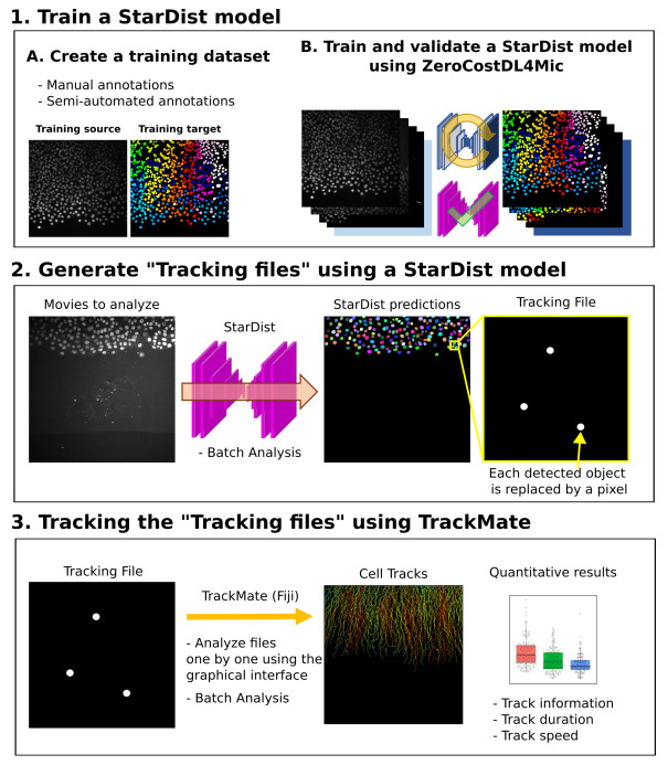
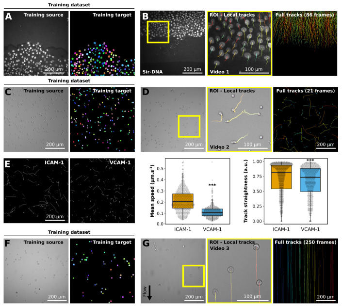
The ability of cells to migrate is a fundamental physiological process involved in embryonic development, tissue homeostasis, immune surveillance, and wound healing. Therefore, the mechanisms governing cellular locomotion have been under intense scrutiny over the last 50 years. One of the main tools of this scrutiny is live-cell quantitative imaging, where researchers image cells over time to study their migration and quantitatively analyze their dynamics by tracking them using the recorded images. Despite the availability of computational tools, manual tracking remains widely used among researchers due to the difficulty setting up robust automated cell tracking and large-scale analysis. Here we provide a detailed analysis pipeline illustrating how the deep learning network StarDist can be combined with the popular tracking software TrackMate to perform 2D automated cell tracking and provide fully quantitative readouts. Our proposed protocol is compatible with both fluorescent and widefield images. It only requires freely available and open-source software (ZeroCostDL4Mic and Fiji), and does not require any coding knowledge from the users, making it a versatile and powerful tool for the field. We demonstrate this pipeline's usability by automatically tracking cancer cells and T cells using fluorescent and brightfield images. Importantly, we provide, as supplementary information, a detailed step-by-step protocol to allow researchers to implement it with their images.

Keywords: Automated tracking; Cell migration; Deep-learning; Image analysis; StarDist; TrackMate.


Conflict of interest statement

No competing interests were disclosed.

Figures

Figure 1.
Figure 1.. Workflow depicting how StarDist and TrackMate can be combined to track cells automatically.
Figure 2.
Figure 2.. Example of datasets analyzed using StarDist and TrackMate.
( A, B) Migration of MCF10DCIS.com, labeled with Sir-DNA, recorded using a spinning disk confocal microscope and automatically tracked. Examples of images used to train StarDist ( A), and an example of results obtained using automated tracking are displayed ( B, Video 1). The yellow square indicates a magnified ROI, where the local track of each nucleus is displayed. The full cell tracks are displayed on the left. Tracks are color-coded as a function of their maximum instantaneous velocity (blue slow, red fast tracks). ( CE) Migration of activated T cell plated on VCAM-1 or ICAM-1, recorded using a brightfield microscope and automatically tracked. Examples of images used to train StarDist ( C) and an example of results obtained using automated tracking are displayed ( D, Video 2). ( E) Comparison of the migration of activated T cells on VCAM-1 or ICAM-1. Track mean speed and track straightness were quantified. Data are displayed as boxplots. *** p-value = <0.001, p-values were determined using a randomization test. ( F, G) Cancer cells flowing in a microfluidic chamber, recorded live using a brightfield microscope and automatically tracked (Video 3). Examples of images used to train StarDist ( F), and an example of results obtained using automated tracking are displayed ( G). The full tracks shown here were color-coded as a function of their x coordinate.

References

    1. Jacquemet G, Baghirov H, Georgiadou M, et al. : L-type calcium channels regulate filopodia stability and cancer cell invasion downstream of integrin signalling. Nat Commun. 2016;7:13297. 10.1038/ncomms13297 - DOI - PMC - PubMed
    1. Jacquemet G, Morgan MR, Byron A, et al. : Rac1 is deactivated at integrin activation sites through an IQGAP1-filamin-A-RacGAP1 pathway. J Cell Sci. 2013;126(Pt 18):4121–4135. 10.1242/jcs.121988 - DOI - PMC - PubMed
    1. DuChez BJ: Automated Tracking of Cell Migration with Rapid Data Analysis. Curr Protoc Cell Biol. 2017;76:12.12.1–12.12.16. 10.1002/cpcb.28 - DOI - PMC - PubMed
    1. Cordelières FP, Petit V, Kumasaka M, et al. : Automated Cell Tracking and Analysis in Phase-Contrast Videos (iTrack4U): Development of Java Software Based on Combined Mean-Shift Processes. PLoS One. 2013;8(11):e81266. 10.1371/journal.pone.0081266 - DOI - PMC - PubMed
    1. McQuin C, Goodman A, Chernyshev V, et al. : CellProfiler 3.0: Next-generation image processing for biology. PLoS Biol. 2018;16(7):e2005970. 10.1371/journal.pbio.2005970 - DOI - PMC - PubMed

Publication types