Electrophysiologic study and catheter ablation of a supraventricular tachycardia in a patient with inferior vena cava congenital anomaly
- PMID: 33224591
- PMCID: PMC7675164
Electrophysiologic study and catheter ablation of a supraventricular tachycardia in a patient with inferior vena cava congenital anomaly
Abstract
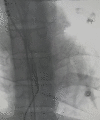
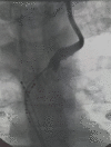
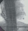
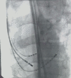
Electrophysiologic procedures are performed widely nowdays, for the successful treatment of several cardiac arrhythmias. In this case report, we describe a rare congenital anomaly of the inferior vena cava, as an incidental finding during a scheduled electrophysiologic procedure for a supraventricular tachycardia ablation. The patient is a 32 year old male with an unremarkable medical history, suffering from sustained episodes of paroxysmal tachycardia. The electrophysiological maneuvers confirmed the presence of atrioventricular nodal reentry tachycardia, followed by a successful slow pathway ablation. We provide imaging details and guidance on the successful catheter positioning. In cases like this, the prognosis is excellent, while the follow up of our patient was unremarkable.
Keywords: Inferior vena cava; congenital anomaly; electrophysiologic study.
AJCD Copyright © 2020.
Conflict of interest statement
None.
Figures


References
-
- Kandpal H, Sharma R, Gamangatti S, Srivastava DN, Vashisht S. Srivastava and sushma vashisht. imaging the inferior vena cava: a road less travelled. Radiographics. 2008;28:669–689. - PubMed
-
- Sheth S, Fishman EK. Imaging of the Inferior Vena Cava with MDCT. AJR Am J Roentgenol. 2007:1243–1251. - PubMed
-
- Bass JE, Redwine MD, Kramer LA, Huynh PT, Harris JH Jr. Spectrum of congenital anomalies of the inferior vena cava: cross-sectional imaging findings. Radiographics. 2000;20:639–652. - PubMed
-
- Kellman GM, Alpern MB, Sandler MA, Craig BM. Computed tomography of vena caval anomalies with embryologic correlation. Radiographics. 1988;8:533–556. - PubMed
-
- Awais M, Rehman A, Baloch NU, Salam B. Multiplanar imaging of inferior vena cava variants. Abdominal Imaging. 2014;40:159–166. - PubMed
Publication types
LinkOut - more resources
Full Text Sources
