. 2020 Mar;29(2):46-62.
doi: 10.1007/s40629-020-00118-6. Epub 2020 Mar 2.

Mast cells and IgE in defense against lethality of venoms: Possible "benefit" of allergy[]

Affiliations

Mast cells and IgE in defense against lethality of venoms: Possible "benefit" of allergy[]

Stephen J Galli et al. Allergo J Int. 2020 Mar.

Abstract

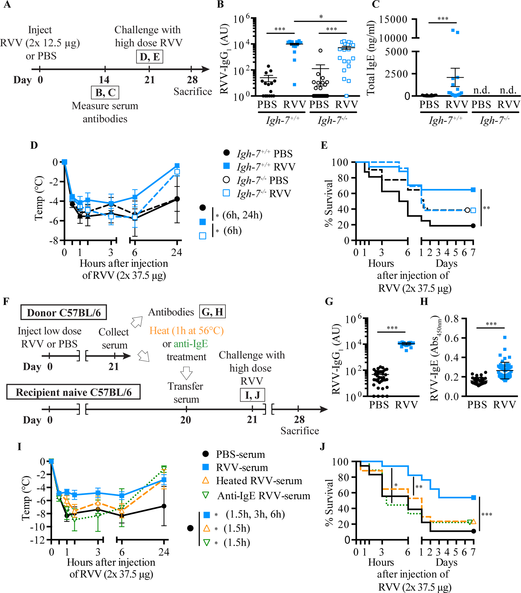
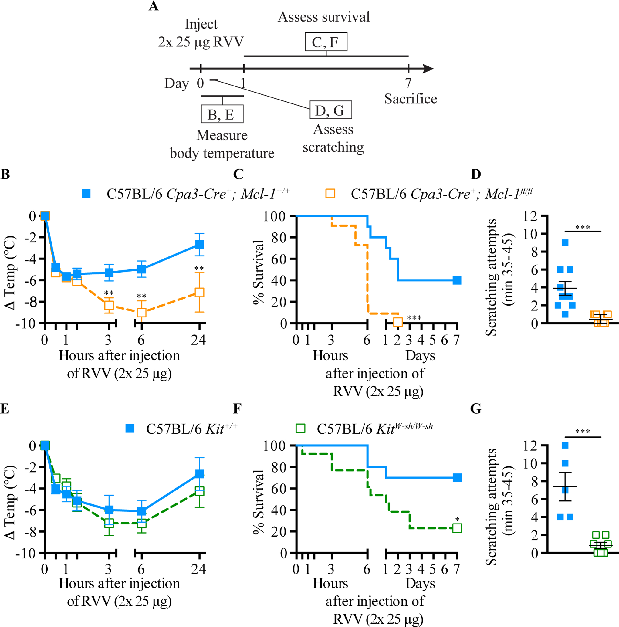
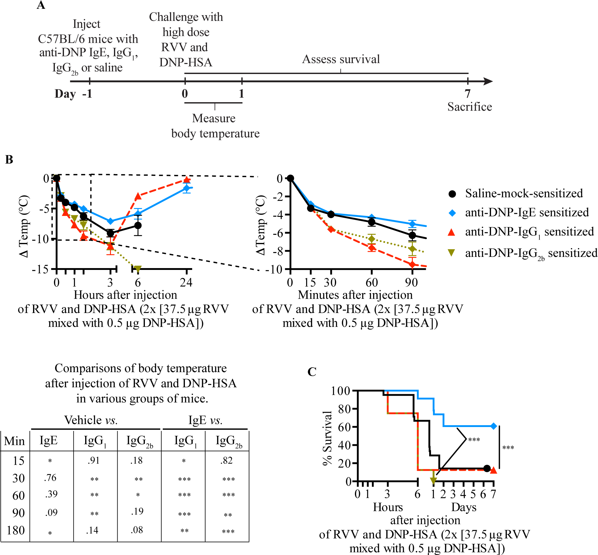
Physicians think of mast cells and IgE primarily in the context of allergic disorders, including fatal anaphylaxis. This 'bad side' of mast cells and IgE is so well accepted that it can be difficult to think of them in other contexts, particularly those in which they may have beneficial functions. However, there is evidence that mast cells and IgE, as well as basophils (circulating granulocytes whose functions partially overlap with those of mast cells), can contribute to host defense as components of adaptive type 2 immune responses to helminths, ticks and certain other parasites. Accordingly, allergies often are conceptualized as "misdirected" type 2 immune responses, in which IgE antibodies are produced against any of a diverse group of apparently harmless antigens, and against components of animal venoms. Indeed, certain unfortunate patients who have become sensitized to venoms develop severe IgE-associated allergic reactions, including fatal anaphylaxis, upon subsequent venom exposure. In this review, we will describe evidence that mast cells can enhance innate resistance, and survival, to challenge with reptile or arthropod venoms during a first exposure to such venoms. We also will discuss findings indicating that, in mice surviving an initial encounter with venom, acquired type 2 immune responses, IgE antibodies, the high affinity IgE receptor (FcεRI), and mast cells can contribute to acquired resistance to the lethal effects of both honeybee venom and Russell's viper venom. These findings support the hypothesis that mast cells and IgE can help protect the host against venoms and perhaps other noxious substances.

Keywords: Allergy; IgE; Th2 cell immunity; toxin hypothesis; venom.


Conflict of interest statement

All authors declare no conflict of interest.

Figures

Fig. 1
Fig. 1. Effects of treatment with anti-basophil serum (ABS), anti-eosinophil serum (AES) or normal rabbit serum (NRS) on feeding success of larval Amblyomma americanum ticks in a second infestation of guinea pigs.
One hundred larval Amblyomma americanum ticks were placed on the flanks of “nonsensitized” (naïve) guinea pigs or guinea pigs that had been “sensitized” 26 days earlier by a primary infestation of A. americanum. The naïve guinea pigs (A) and one group of sensitized hosts (B) received no serum, other sensitized animals were treated with NRS (C), ABS (D), or AES (E), as described in [45]. The number (Tick Yield, left) and weight (Tick Weight, right) of engorged ticks was determined at 90 h of infestation. ABS completely ablated immunity; AES partially impaired resistance; NRS had no effect. Data (mean +/− SE) were pooled from three separate experiments, with the total number of animals in each group shown in parentheses. NS = not significant (P > 0.05). Differences among the experimental groups were analyzed by the Newman-Keuls multiple sample comparison test. [This is a modified version of Fig. 2 in Brown SJ, Galli SJ, Gleich GJ, Askenase PW: Ablation of immunity to Amblyomma americanum by anti-basophil serum: cooperation between basophils and eosinophils in expression of immunity to ectoparasites (ticks) in guinea pigs. J Immunol 1982;129:790–6 (ref. [45]) reprinted with the permission of the publisher. Copyright 1982. The American Association of Immunologists, Inc.]
Fig. 2
Fig. 2. Making “mast cell knock-in mice”.
(1) Mast cells can be generated from bone marrow cells (or other hematopoietic cells; e.g., those in the fetal liver) from wild type mice or from mutant or transgenic mice with specific genetic alterations of interest [–63]. Alternatively, (2) embryonic stem (ES) cell-derived cultured mast cells (ESCMCs) can be generated from wild type or genetically altered ES cells [64, 65], or (3) various mast cell populations can be transduced in vitro with shRNA to diminish expression of specific genes of interest [15, 66]. (4) Such bone marrow-, ES cell-, [or fetal liver-] derived cultured mast cells, or shRNA-transduced mast cells, can then be transplanted into mast cell-deficient c-kit mutant mice, such as WBB6F1-KitW/KitW-v mice [63, 67] or C57BL/6-KitW-sh/KitW-sh mice [68, 69], or into C57BL/6-Cpa3-Cre;Mcl-1fl/fl mice [70] (which we informally refer to as “Hello Kitty mice”, which have wild type c-kit), to produce mast cell knock-in mice. Note: BMCMCs can be injected into genetically mast cell-deficient mice intravenously (i.v.), intraperitoneally (i.p.), or intradermally (i.d.), or into the joints or meninges, etc., but there is a more limited experience with the engraftment of other types of MCs, such as EMCMCs, than with BMCMCs. (5) A suitable interval is then allowed for engraftment and phenotypic “maturation” of the adoptively-transferred mast cells (the length of this interval can be varied based on the route of mast cell transfer, the anatomical site of interest, the particular biological response being analyzed, etc.). The importance of mast cell function(s) in biological responses can be analyzed by comparison of the responses in the appropriate wild type or littermate control mice (6), the corresponding mutant mast cell-deficient mice (7), and selectively mast cell-engrafted mutant mice (mast cell knock-in mice) (8). The contributions of specific mast cell products (surface structures, signaling molecules, secreted products, and so on) to such biological responses can be analyzed by comparing the features of the responses of interest in mast cell knock-in mice engrafted with wild-type mast cells versus mast cells derived from mice or ES cells that lack or express genetically altered forms of such products or that have been transduced with shRNA to silence the specific genes that encode these products. An important part of the analysis of the mast cell knock-in mice used in particular experiments is to assess the numbers and anatomic distribution, and, for certain experiments, aspects of the phenotype, of the adoptively-transferred mast cells, as, depending on the type of in vitro-derived mast cells used, the route of administration, and other factors, these may differ from those of the corresponding native populations of mast cells in the corresponding wild type mice [14, 71, 72]. [This is a modified version of Fig. 2 in Metz M, Grimbaldeston MA, Nakae S, Piliponsky AM, Tsai M, Galli SJ. Mast cells in the promotion and limitation of chronic inflammation. Immunol Rev 2007; 217:304–28 (ref. [71]), reprinted with the permission of the publisher, John Wiley and Sons.]
Fig. 3
Fig. 3. Mast cells can diminish Heloderma suspectum venom (H.s.v.)-induced hypothermia and mortality through MCP4-dependent mechanisms.
Changes in rectal temperatures after i.d. injection of H.s.v. (25 μg in 20 μl DMEM solution) into the ear pinnae (one ear pinna of each mouse) of: (A) WT WBB6F1-Kit+/+, mast cell-deficient WBB6F1-KitW/W-v, and WT BMCMCs→KitW/W-v mice (i.e., WBB6F1-KitW/W-v mice which had been engrafted, 6–8 weeks before venom challenge, in one ear pinna with 2 million BMCMCs derived from WT WBB6F1-Kit+/+ mice) (the death rates of Kit+/+, WT BMCMCs→KitW/W-v, and KitW/W-v mice within 24 h after H.s.v. injection were 0% [0/21], 7% [1/15, P = 0.42 vs. Kit+/+ mice], and 65% [13/20, P < 0.0001 vs. Kit+/+ mice], respectively); (B) WT C57BL/6-Kit+/+, mast cell-deficient C57BL/6-KitW-sh/W-sh, WT BMCMCs→KitW-sh/W-sh, and Mcpt4−/− BMCMCs→KitW-sh/W-sh mice (the death rates of Kit+/+, WT BMCMCs→KitW-sh/W-sh, Mcpt4−/− BMCMCs→KitW-sh/W-sh, and KitW-sh/W-sh mice within 24 h after H.s.v. injection were 5% [1/19], 11% [2/18, P = 0.48 vs. Kit+/+ mice], 43% [6/14, P = 0.01 vs. Kit+/+ mice], and 50% [10/20, P = 0.006 vs. Kit+/+ mice], respectively); or (C) WT C57BL/6-Kit+/+ mice, C57BL/6-Cpa3Y356L,E378A mice (which have a catalytically inactive CPA3) and C57BL/6-Mcpt4−/− mice (the death rates of Kit+/+, Cpa3Y356L,E378A, and Mcpt4−/− mice within 24 h after H.s.v. injection were 7% [1/15], 0% [0/14, P = 0.52 vs. Kit+/+ mice], 40% [6/15, P = 0.007 vs. Kit+/+ mice], respectively). Each figure shows data pooled from at least three independent experiments with each group of mice (n = 2–5 mice per group per each individual experiment). **P < 0.01, ***P < 0.001 versus WT WBB6F1-Kit+/+ or WT C57BL/6-Kit+/+ mice; P < 0.01~0.001 versus each other group (A-C). (D) Extensive degranulation of mast cells (some indicated by closed arrowheads) 1 h after i.d. injection of H.s.v. (25 μg in 20 μl DMEM), but not vehicle (DMEM) alone (mast cells without evidence of degranulation are indicated by open arrowheads) in WT C57BL/6 mice (Toluidine blue stain; scale bar: 50 micrometers). (E) Degranulation of mast cells 60 min after i.d. injection of H.s.v. (25 μg in 20 μl DMEM) or vehicle (DMEM) alone in WT C57BL/6, Mcpt4−/−, or Cpa3Y356L,E378A mice (injection was into one ear pinna of each mouse). ***P < 0.001 versus corresponding vehicle-injected groups; NS = not significant (P > 0.05) versus values for WT mice. [This is a reproduction of Fig. 1 from Akahoshi M, Song CH, Piliponsky AM, Metz M, Guzzetta A, Abrink M, Schlenner SM, Feyerabend TB, Rodewald HR, Pejler G, Tsai M, Galli SJ. Mast cell chymase reduces the toxicity of Gila monster venom, scorpion venom, and vasoactive intestinal polypeptide in mice. J Clin Invest 2011;121:4180–91 (ref. [18]), reprinted with the permission of the publisher, the American Society for Clinical Investigation.]
Fig. 4
Fig. 4. Mast cells can enhance innate resistance to high levels of endogenous peptides and structurally similar peptides in reptile venoms.
Mast cell cytoplasmic granules contain proteases such as carboxypeptidase A3 (CPA3 [mCPA3 = mouse CPA3]) and mast cell protease 4 (MCP4 [mMCP4 = mouse MCP4]) that, upon secretion by activated mast cells, can degrade certain endogenous peptides, such as endothelin-1 (ET-1) and vasoactive intestinal polypeptide (VIP), respectively, as well as structurally similar peptides contained in the venoms of poisonous reptiles, such as sarafotoxin 6b in the venom of the Israeli mole viper (Atractaspis engaddensis) and helodermin in the venom of the Gila monster (Heloderma suspectum). The ability of mast cells to be activated to degranulate by components of venoms such as these, which can act at the same receptors which recognize the corresponding structurally similar endogenous peptides, permits mast cells to release proteases that can reduce the toxicity of these peptides and which help to enhance the survival of mice injected with the whole venoms of these reptiles, that contain many toxins in addition to sarafotoxin 6b and helodermin. This mechanism may also permit mast cells to restore homeostasis in settings associated with markedly increase levels of the endogenous peptides. [This is a reproduction of Fig. 4 from Galli SJ. The 2014 Rous-Whipple Award Lecture. The mast cell-IgE paradox: From homeostasis to anaphylaxis. Am J Pathol 2016;186:212–24 (ref. [42]), reprinted with the permission of the publisher, Elsevier for the American Society for Investigative Pathology.]
Fig. 5
Fig. 5. Evidence that IgE antibodies contribute to acquired enhanced resistance to the toxicity and lethality of Russell’s viper venom.
A. Outline of experiments with IgE-deficient (Igh-7−/−) and control (Igh-7+/+) C57BL/6 mice (B-E). B, C. Serum RVV-specific IgG1 (B) and total IgE (C). D, E. Body temperature (D) and survival (E). F. Outline of serum transfer experiments in C57BL/6 mice (G-J). G, H. Serum RVV-specific IgG1 (G) and total IgE (H). I, J. Body temperature (I) and survival (J). Data were pooled from 3–4 experiments (n= 9–25/group). P values: Mann-Whitney test (B, C, G, H), Student’s t test (D, I) and Mantel-Cox test (E, J). [This is a reproduction of Fig. 3 from Starkl P, Marichal T, Gaudenzio N, Reber LL, Sibilano R, Tsai M, Galli SJ. IgE antibodies, FcεRIα, and IgE-mediated local anaphylaxis can limit snake venom toxicity. J Allergy Clin Immunol 2016;137:246–57 (ref. [105]), reprinted with the permission of the publisher, Elsevier.]
Fig. 6
Fig. 6. Evidence that mast cells contribute to innate resistance to the toxicity and lethality of Russell’s viper venom, as well as to behavioral responses to envenomation.
A. Experimental outline. B and E, body temperature; C and F, survival; D and G, scratching attempts, of mast cell-deficient Cpa3-Cre+; Mcl-1fl/fl (B-D) and KitW-sh/W-sh (E-G) mice and corresponding control mice after RVV injection. P values: (B, D, E, G) Student’s t test; (C, F) Mantel-Cox test. Data were pooled from 2–4 experiments (n=5–21/group). [This is a reproduction of Fig. 2 of Starkl P, Marichal T, Gaudenzio N, Reber LL, Sibilano R, Tsai M, Galli SJ. IgE antibodies, FcεRIα and IgE-mediated local anaphylaxis can limit snake venom toxicity. J Allergy Clin Immunol 2016;137:246–57 (ref. [105]), reprinted with the permission of the publisher, Elsevier.]
Fig. 7
Fig. 7. IgE-dependent local mast cell activation induced by activation with a single antigen can enhance resistance to the lethality of Russell’s viper venom.
A. Experimental outline. B, C. Body temperature (B) and survival (C) of C57BL/6 mice treated with 3 s.c. injections of saline alone or containing 50 ng anti-DNP IgE, IgG1 or IgG2b antibody and challenged 18 h later with 2 s.c. injections, each containing 37.5 μg RVV and 0.5 μg DNP-HSA. Data were pooled from 2–5 independent experiments (n=10–25/group). P values: Student’s t test (B); Mantel-Cox test (C). [This is a reproduction of Fig. 5 from Starkl P, Marichal T, Gaudenzio N, Reber LL, Sibilano R, Tsai M, Galli SJ. IgE antibodies, FcεRIα and IgE-mediated local anaphylaxis can limit snake venom toxicity. J Allergy Clin Immunol 2016;137:246–57 (ref. [105]), reprinted with the permission of the publisher, Elsevier.]

Similar articles

Cited by

References

    1. Pawankar R, Canonica GW, Holgate ST, Lockey RF. Allergic diseases and asthma: a major global health concern. Curr Opin Allergy Clin Immunol 2012; 12:39–41. - PubMed
    1. Paul WE, Zhu J. How are TH2-type immune responses initiated and amplified? Nat Rev Immunol 2010;10:225–35. - PMC - PubMed
    1. Galli SJ, Tsai M. IgE and mast cells in allergic disease. Nat Med 2012;18:693–704. - PMC - PubMed
    1. Pulendran B, Artis D. New paradigms in type 2 immunity. Science 2012;337:431–5. - PMC - PubMed
    1. Kinet JP. The high-affinity IgE receptor (FcƐRI): from physiology to pathology. Annu Rev Immunol 1999;17:931–72. - PubMed