

When to use intravascular ultrasound or optical coherence tomography during percutaneous coronary intervention?
- PMID: 33224766
- PMCID: PMC7666918
- DOI: 10.21037/cdt-20-206
When to use intravascular ultrasound or optical coherence tomography during percutaneous coronary intervention?
Abstract
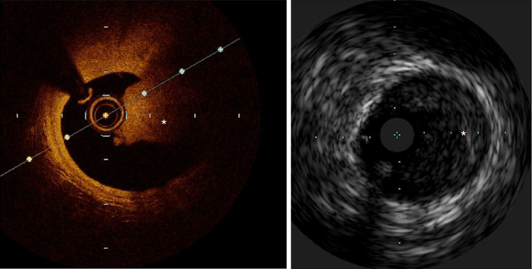
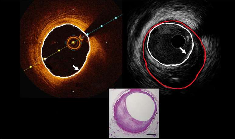
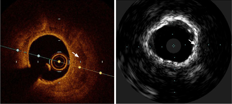
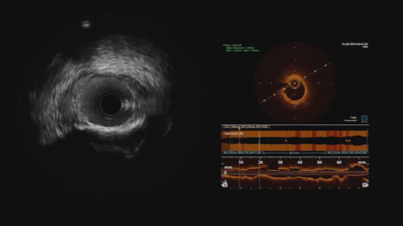
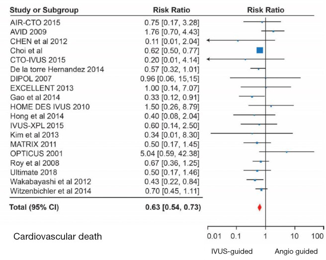
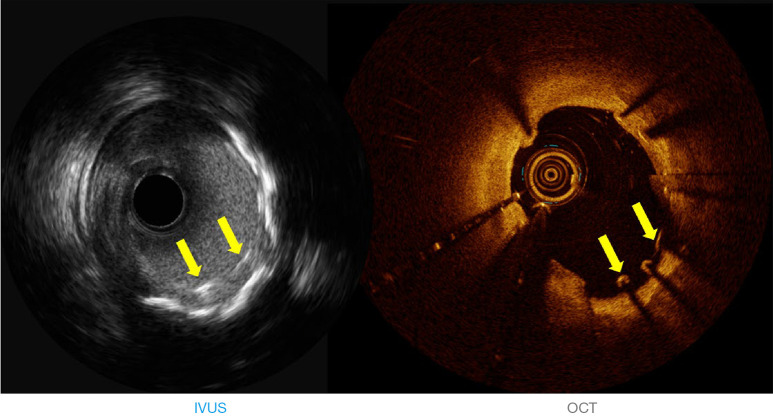
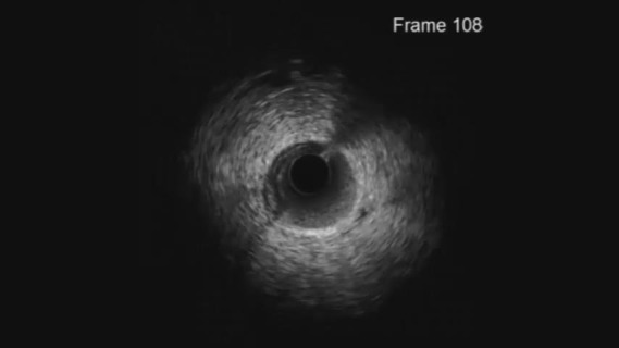
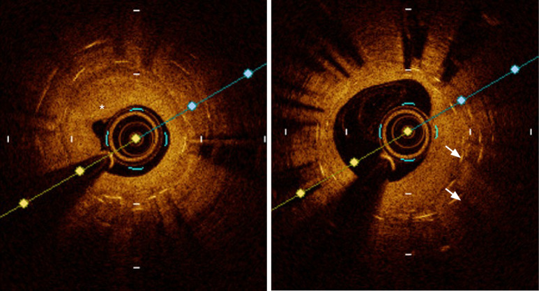
Intravascular ultrasound (IVUS) and optical coherence tomography (OCT) are intravascular imaging technologies widely used in the cardiac catheterization laboratory. The impact of these modalities for optimizing the acute and longer-term clinical impact following percutaneous coronary intervention (PCI) is supported by a wealth of clinical evidence. Intravascular imaging provides unique information for enhanced lesion preparation, optimal stent sizing, recognizing post PCI complications, and the etiology of stent failure. This review compares and contrasts the key aspects of these imaging modalities during PCI.
Keywords: Intravascular ultrasound (IVUS); cardiovascular mortality; optical coherence tomography (OCT); percutaneous coronary intervention (PCI); repeat revascularization.
2020 Cardiovascular Diagnosis and Therapy. All rights reserved.
Conflict of interest statement
Conflicts of Interest: All authors have completed the ICMJE uniform disclosure forms (available at http://dx.doi.org/10.21037/cdt-20-206). The series “Intracoronary Imaging” was commissioned by the editorial office without any funding or sponsorship. The authors have no other conflicts of interest to declare.
Figures
References
-
- Lee MS, Shlofmitz E, Kong J, et al. Impact of the Use of Intravascular Imaging on Patients Who Underwent Orbital Atherectomy. J Invasive Cardiol 2018;30:77-80. - PubMed
Publication types
LinkOut - more resources
Full Text Sources
Miscellaneous