Review
. 2020 Oct;10(5):1580-1603.
doi: 10.21037/cdt-20-479.

Mechanics of right ventricular dysfunction in pulmonary arterial hypertension and heart failure with preserved ejection fraction

Affiliations
Review

Mechanics of right ventricular dysfunction in pulmonary arterial hypertension and heart failure with preserved ejection fraction

Roberto J Bernardo et al. Cardiovasc Diagn Ther. 2020 Oct.

Abstract

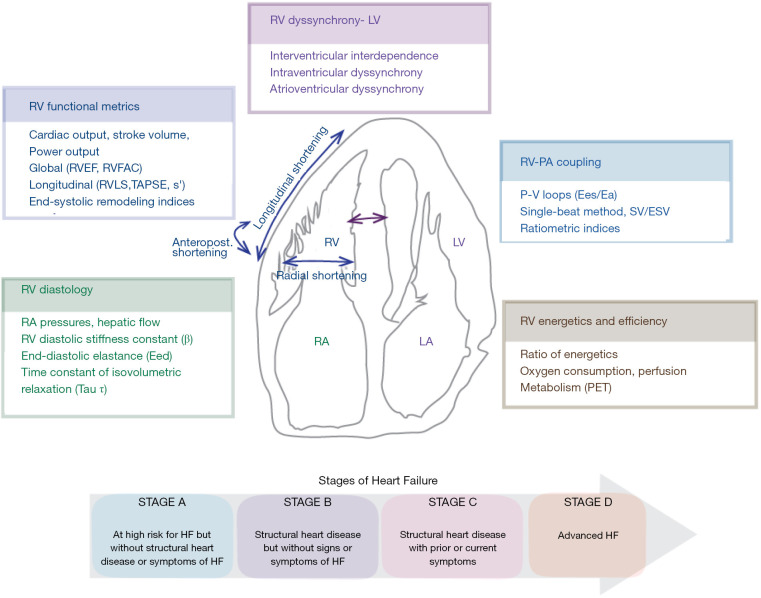
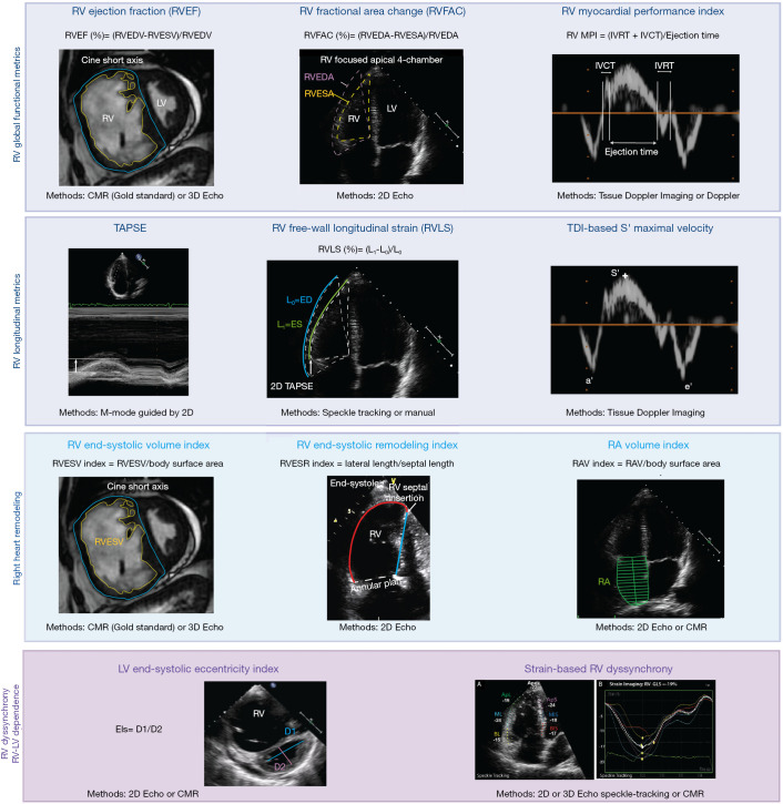
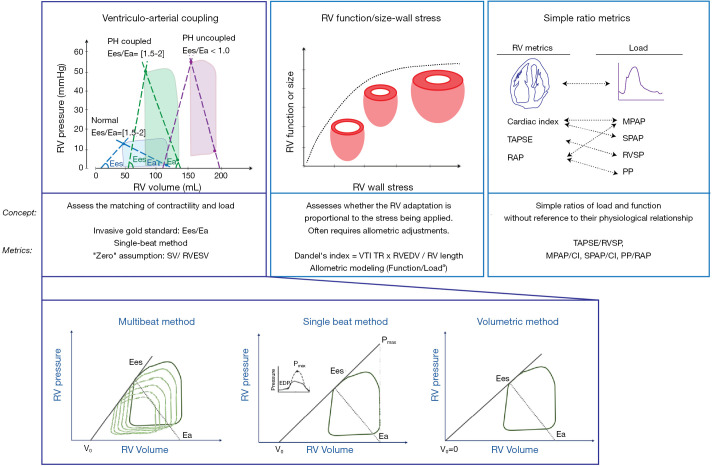
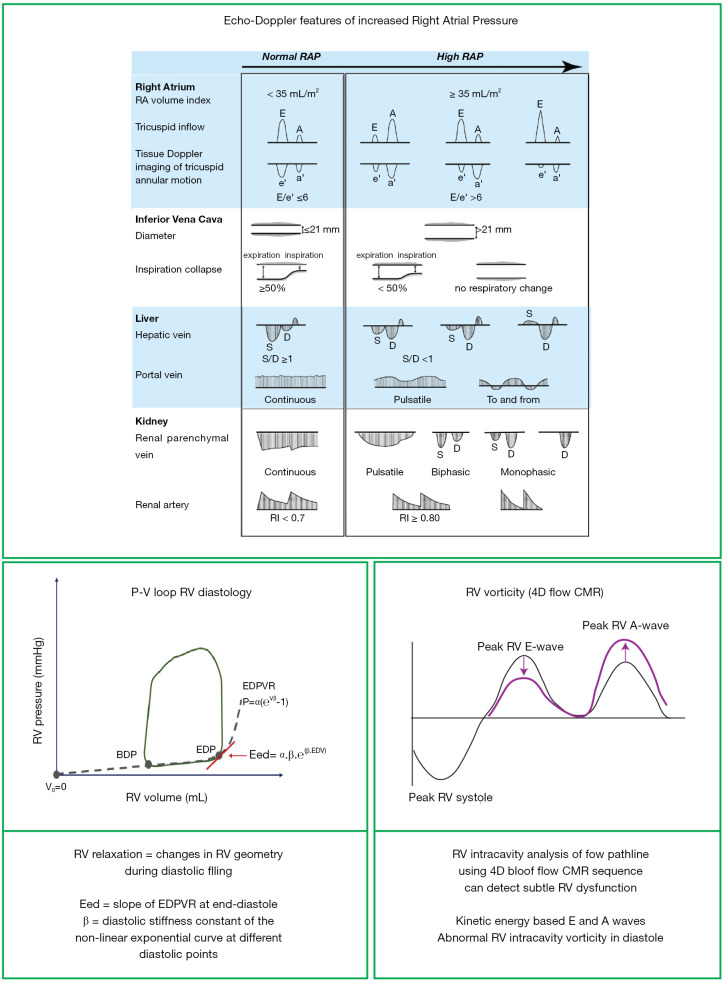
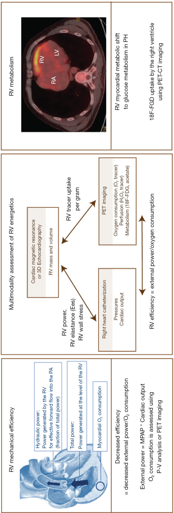
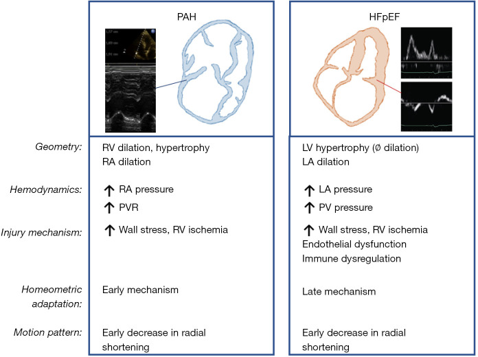
Right ventricular (RV) dysfunction is the most important determinant of survival in patients with pulmonary hypertension (PH). The manifestations of RV dysfunction not only include changes in global RV systolic function but also abnormalities in the pattern of contraction and synchrony. The effects of PH on the right ventricle have been mainly studied in patients with pulmonary arterial hypertension (PAH). However, with the demographic shift towards an aging population, heart failure with preserved ejection fraction (HFpEF) has become an important etiology of PH in recent years. There are significant differences in RV mechanics, function and adaptation between patients with PAH and HFpEF (with or without PH), which are related to different patterns of remodeling and dysfunction. Due to the unique features of the RV chamber, its connection with the main pulmonary artery and the pulmonary circulation, an understanding of the mechanics of RV function and its clinical significance is mandatory for both entities. In this review, we describe the mechanics of the pressure overloaded right ventricle. We review the different mechanical components of RV dysfunction and ventricular dyssynchrony, followed by insights via analysis of pressure-volume loop, energetics and novel blood flow patterns, such as vortex imaging. We conduct an in-depth comparison of prevalence and characteristics of RV dysfunction in HFpEF and PAH, and summarize key outcome studies. Finally, we provide a perspective on needed and expected future work in the field of RV mechanics.

Keywords: Heart failure with preserved ejection fraction (HFpEF); mechanical efficiency; pulmonary hypertension (PH); right ventricular dysfunction (RV dysfunction); ventriculo-arterial coupling.


Conflict of interest statement

Conflicts of Interest: All authors have completed the ICMJE uniform disclosure form (available at http://dx.doi.org/10.21037/cdt-20-479). The series “Right Ventricular Dysfunction” was commissioned by the editorial office without any funding or sponsorship. GH served as the unpaid Guest Editor of the series. GH reports grants from German Research Foundation (DFG; HA4348/2-2 and HA4348/6-2 KFO311), from Federal Ministry of Education and Research (BMBF ViP+ program-03VP08053; BMBF 01KC2001B), from European Pediatric Pulmonary Vascular Disease Network (www.pvdnetwork.org), outside the submitted work. AYD reports grants from Edwards (2019) research (equipment grant), outside the submitted work; and Speaker bureau and KOL for CAE Healthcare and Masimo. Dr. FSdM reports grants from The Netherlands CardioVascular Research Initiative: the Dutch Heart Foundation, Dutch Federation of University Medical Centres, the Netherlands Organisation for Health Research and Development, and the Royal Netherlands Academy of Sciences (CVON-2012-08 PHAEDRA, CVON-2018-29 PHAEDRA-IMPACT, CVON-2017-10 Dolphin-Genesis), from The Netherlands Organization for Scientific Research (NWO-VIDI: 917.18.338), from The Dutch Heart Foundation Dekker senior post-doc grant (2018T059), outside the submitted work. The authors have no other conflicts of interest to declare.

Figures

Figure 1
Figure 1
Graphical abstract. Components of right ventricular (RV) mechanics. ESV, end-systolic volume; HF, heart failure; LA, left atrium; LV, left ventricle; PET, positron emission tomography; P-V loops, pressure-volume loops; RA, right atrium; RV, right ventricle; RVEF, RV ejection fraction; RVFAC, RV fractional area change; RVLS, RV longitudinal strain; TAPSE, tricuspid annular plane systolic excursion; S’, Tissue-Doppler tricuspid annular S’ wave maximal velocity; SV, stroke volume.
Figure 2
Figure 2
Right ventricular clinical imaging metrics. 2D Echo, two-dimensional echocardiography; 3D, three-dimensional echocardiography; CMR, cardiac magnetic resonance imaging; EIs, end-systolic eccentricity index; IVCT, isovolumetric contraction time; IVRT, isovolumetric relaxation time; LV, left ventricular; RA, right atrium; RV, right ventricular; TAPSE, tricuspid annular plane systolic excursion; TDI, tissue doppler imaging.
Figure 3
Figure 3
Right ventricular-pulmonary arterial coupling. Upper panels (left to right): pressure-volume loop analysis and estimation of ventriculo-arterial coupling; correlation between RV wall stress and RV dilation/size and representation of different ratio metric indices of RV load and function. Lower panels (left to right): Methods for estimation of ventriculo-arterial coupling. The multibeat method requires obtaining different sets of pressure-volume loops at decreasing levels of preload (left). In the single beat method, the RV pressure waveform tracing is used for estimation of Pmax (center). In the volumetric method (right), V0 is assumed to be equal to zero, and Ees is estimated by the ratio of end-systolic pressure to end-systolic volume.
Figure 4
Figure 4
Right ventricular (RV) diastology. BDP, beginning of diastolic pressure; CMR, cardiac magnetic resonance; Eed, ventricular end-diastolic elastance; EDP, end-diastolic pressure; EDPVR, end-diastolic pressure-volume relationship; P-V, pressure-volume loops; V0, Volume intercept at zero pressure. Adapted with permission from Tang et al.
Figure 5
Figure 5
Right ventricular (RV) efficiency and energetics. CT, computed tomography; MPAP, mean pulmonary arterial pressure; P-V, pressure-volume loops analysis; PET, positrons emission tomography; PH, pulmonary hypertension.
Figure 6
Figure 6
Schematic comparison of RV mechanics in PAH vs. HFpEF-PH. HFpEF, heart failure with preserved ejection fraction; LA, left atrial; LV, left ventricular; PAH, pulmonary arterial hypertension; PV, pulmonary vein; PVR, pulmonary vascular resistance; RA, right atrial; RV, right ventricular.

References

    1. Champion HC, Michelakis ED, Hassoun PM. Comprehensive invasive and noninvasive approach to the right ventricle-pulmonary circulation unit: state of the art and clinical and research implications. Circulation 2009;120:992-1007. 10.1161/CIRCULATIONAHA.106.674028 - DOI - PubMed
    1. Vanderpool RR, Saul M, Nouraie M, et al. Association Between Hemodynamic Markers of Pulmonary Hypertension and Outcomes in Heart Failure With Preserved Ejection Fraction. JAMA Cardiol 2018;3:298-306. 10.1001/jamacardio.2018.0128 - DOI - PMC - PubMed
    1. Vonk Noordegraaf A, Chin KM, Haddad F, et al. Pathophysiology of the right ventricle and of the pulmonary circulation in pulmonary hypertension: an update. Eur Respir J 2019;53:1801900. 10.1183/13993003.01900-2018 - DOI - PMC - PubMed
    1. Hoeper MM, Humbert M, Souza R, et al. A global view of pulmonary hypertension. Lancet Respir Med 2016;4:306-22. 10.1016/S2213-2600(15)00543-3 - DOI - PubMed
    1. Rich S, Pogoriler J, Husain AN, et al. Long-term effects of epoprostenol on the pulmonary vasculature in idiopathic pulmonary arterial hypertension. Chest 2010;138:1234-9. 10.1378/chest.09-2815 - DOI - PMC - PubMed