Review
. 2020 Oct;10(5):1646-1658.
doi: 10.21037/cdt-20-450.

Right ventricular dysfunction and long-term risk of death

Affiliations
Review

Right ventricular dysfunction and long-term risk of death

Jason L Sanders et al. Cardiovasc Diagn Ther. 2020 Oct.

Abstract

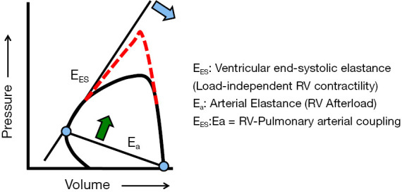
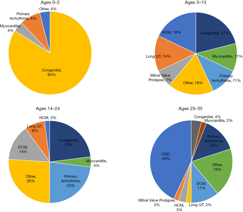
Sudden cardiac death (SCD), or sudden loss of life-sustaining systemic and cerebral perfusion, is most often due to left ventricular (LV) dysfunction secondary to ischemic or structural cardiac disease or channelopathies. Degeneration of sinus rhythm into ventricular tachycardia and ultimately ventricular fibrillation is the final common pathway for most heart failure patients. Right ventricular (RV) dysfunction is recognized as an independent contributor to worsening heart failure. There is emerging evidence that RV dysfunction may also be an independent predictor of SCD. This review examines the role of RV dysfunction on modifying long term risk of SCD, and explores possible mechanisms that may underlie SCD. The RV has unique anatomy and physiology compared to the LV. Subsequently, we begin with a review of cardiac embryology, focusing on the chambers, valves, coronary arteries, and cardiac conduction system to understand the origins of RV dysfunction. Static and dynamic physiology of the RV is contrasted with that of the LV. Particular emphasis is placed on ventriculo-arterial coupling, mechanical cardiac constraint, and ventricular interdependence. The epidemiology of SCD is briefly reviewed to highlight how causes of SCD are age-specific. In turn, the age-specific causes of RV dysfunction are presented, including those which predominate in childhood and adolescence [arrhythmogenic RV dysplasia (ARVD) and hypertrophic cardiomyopathy (HCM)] and older adulthood (cardiac ischemia, chronic congestive heart failure and post-capillary pulmonary hypertension, and pulmonary hypertension). There is a clear need for additional studies on the independent contribution of RV dysfunction to overall functional capacity, SCD-associated mortality, and non-SCD-associated mortality. Discovery would be aided by the development of prospective cohorts with excellent RV phenotyping, coupled with deeper biologic measurements linking mechanisms to clinically relevant outcomes.

Keywords: Right ventricle; arrhythmogenic right ventricular dysplasia (ARVD); hypertrophic cardiomyopathy (HCM); pulmonary hypertension; sudden cardiac death (SCD).


Conflict of interest statement

Conflicts of Interest: The authors have completed the ICMJE uniform disclosure form (available at http://dx.doi.org/10.21037/cdt-20-450). The series “Right Ventricular Dysfunction” was commissioned by the editorial office without any funding or sponsorship. Dr. MK served as the unpaid Guest Editors of the series. Dr. SR reports personal fees and other from Abbott, grants and personal fees from Actelion, personal fees from Acceleron, grants and personal fees from AstraZeneca, grants and personal fees from Bayer, personal fees from BMS, personal fees from Janssen, personal fees from Merck, grants and personal fees from Novartis, grants and personal fees from Pfizer, grants and personal fees from United Therapeutics, personal fees from Vifor, outside the submitted work. Dr. BAM reports other from Acetelion, during the conduct of the study. The author has no other conflicts of interest to declare.

Figures

Figure 1
Figure 1
Systemic consequences of pulmonary hypertension and right heart failure in multiple organ systems. Reproduced with permission from (1).
Figure 2
Figure 2
Three-dimensional characterization of an anomalous right upper pulmonary vein (RUPV)-superior vena cava (SVC) communication. A 67-year-old man presented for the first time with unexplained dyspnea. Advanced imaging indicated that this was due to undiagnosed congenital heart disease causing anomalous pulmonary venous return to the superior vena cava. (A) Still frame image acquired during 3-dimensional transesophageal echocardiography demonstrates contiguous blood flow between the right upper pulmonary vein (outlined by arrowheads) and SVC. Asterisks (*) designate the margins of the anomalous channel between these structures. (B) Multislice 3-dimensional reconstructive computed tomographic angiography (acquired at right anterior oblique of 84°, caudal of 31°) reveals an abnormal RUPV-SVC communication (provided at increased magnification in the inset) with normal insertion of the RUPV into the left atrium (LA). (C) Multislice 3-dimensional reconstructed computed tomographic angiography shows a superior course of the anomalous RUPV from the lung parenchyma without meandering before communication with the SVC. Asterisk (*) indicates RUPV. IVC indicates inferior vena cava; and RA, right atrium. Reproduced with permission from Clarke et al., Circ Cardiovasc Imaging 2013;6(2):349-351.
Figure 3
Figure 3
Right ventricular-pulmonary vascular coupling. The pressure-volume diagram is used to calculate right ventricular (RV)-pulmonary vascular coupling. The pressure-volume diagram allows for the determination of RV end-systolic elastance (EES). EES is unaffected by changes to RV afterload (dashed line) and, therefore, is the best possible load-independent measurement of contractility. By contrast, arterial elastance (Ea) is proportional to pulmonary vascular resistance and is a measurement of RV afterload. The ratio of EES to Ea is a measure of the coupling of the respective ventricular and arterial loads. Thus, a decrease in EES (blue arrow) or an increase in Ea (green arrow) due to elevations in pulmonary vascular resistance, for example, disrupts normal coupling. Reproduced with permission from Maron et al., Pulm Circ 2014;4(4):705-716.
Figure 4
Figure 4
Causes of sudden cardiac arrest by age group. CAD, coronary artery disease; DCM, dilated cardiomyopathy; HCM, hypertrophic cardiomyopathy. Reproduced with permission from Benjamin EJ, Virani SS, Callaway CW, et al. Heart Disease and Stroke Statistics-2018 Update: A Report From the American Heart Association. Circulation 2018;137(12):e67-e492 (16).

References

    1. Rosenkranz S, Howard LS, Gomberg-Maitland M, et al. Systemic Consequences of Pulmonary Hypertension and Right-Sided Heart Failure. Circulation 2020;141:678-93. 10.1161/CIRCULATIONAHA.116.022362 - DOI - PubMed
    1. Kloesel B, DiNardo JA, Body SC. Cardiac Embryology and Molecular Mechanisms of Congenital Heart Disease: A Primer for Anesthesiologists. Anesth Analg 2016;123:551-69. 10.1213/ANE.0000000000001451 - DOI - PMC - PubMed
    1. Emmanouilides GC, Moss AJ, Duffie ER, Jr, et al. Pulmonary Arterial Pressure Changes in Human Newborn Infants from Birth to 3 Days of Age. J Pediatr 1964;65:327-33. 10.1016/S0022-3476(64)80395-4 - DOI - PubMed
    1. Dell'Italia LJ. The right ventricle: anatomy, physiology, and clinical importance. Curr Probl Cardiol 1991;16:653-720. 10.1016/0146-2806(91)90009-Y - DOI - PubMed
    1. Sheehan F, Redington A. The right ventricle: anatomy, physiology and clinical imaging. Heart 2008;94:1510-5. 10.1136/hrt.2007.132779 - DOI - PubMed