

Cardiac multi-scale investigation of the right and left ventricle ex vivo: a review
- PMID: 33224784
- PMCID: PMC7666925
- DOI: 10.21037/cdt-20-269
Cardiac multi-scale investigation of the right and left ventricle ex vivo: a review
Abstract
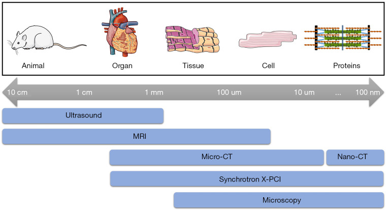
The heart is a complex multi-scale system composed of components integrated at the subcellular, cellular, tissue and organ levels. The myocytes, the contractile elements of the heart, form a complex three-dimensional (3D) network which enables propagation of the electrical signal that triggers the contraction to efficiently pump blood towards the whole body. Cardiovascular diseases (CVDs), a major cause of mortality in developed countries, often lead to cardiovascular remodeling affecting cardiac structure and function at all scales, from myocytes and their surrounding collagen matrix to the 3D organization of the whole heart. As yet, there is no consensus as to how the myocytes are arranged and packed within their connective tissue matrix, nor how best to image them at multiple scales. Cardiovascular imaging is routinely used to investigate cardiac structure and function as well as for the evaluation of cardiac remodeling in CVDs. For a complete understanding of the relationship between structural remodeling and cardiac dysfunction in CVDs, multi-scale imaging approaches are necessary to achieve a detailed description of ventricular architecture along with cardiac function. In this context, ventricular architecture has been extensively studied using a wide variety of imaging techniques: ultrasound (US), optical coherence tomography (OCT), microscopy (confocal, episcopic, light sheet, polarized light), magnetic resonance imaging (MRI), micro-computed tomography (micro-CT) and, more recently, synchrotron X-ray phase contrast imaging (SR X-PCI). Each of these techniques have their own set of strengths and weaknesses, relating to sample size, preparation, resolution, 2D/3D capabilities, use of contrast agents and possibility of performing together with in vivo studies. Therefore, the combination of different imaging techniques to investigate the same sample, thus taking advantage of the strengths of each method, could help us to extract the maximum information about ventricular architecture and function. In this review, we provide an overview of available and emerging cardiovascular imaging techniques for assessing myocardial architecture ex vivo and discuss their utility in being able to quantify cardiac remodeling, in CVDs, from myocyte to whole organ.
Keywords: Ventricular architecture; cardiomyocytes; cardiovascular imaging; multi-scale imaging; myocardium.
2020 Cardiovascular Diagnosis and Therapy. All rights reserved.
Conflict of interest statement
Conflicts of Interest: All authors have completed the ICMJE uniform disclosure form (available at http://dx.doi.org/10.21037/cdt-20-269). The series “Right Ventricular Dysfunction” was commissioned by the editorial office without any funding or sponsorship. The authors have no other conflicts of interest to declare.
Figures
References
-
- LeGrice I, Pope A, Smaill B. The architecture of the heart: myocyte organization and the cardiac extracellular matrix. In: Villarreal FJ. Interstitial fibrosis in heart failure. New York: Springer, 2005:3-21.
-
- LeGrice IJ, Smaill BH, Chai LZ, et al. Laminar structure of the heart: ventricular myocyte arrangement and connective tissue architecture in the dog. Am J Physiol 1995;269:H571-82. - PubMed
Publication types
LinkOut - more resources
Full Text Sources
Research Materials
Miscellaneous