

Metabolic Compartmentalization at the Leading Edge of Metastatic Cancer Cells
- PMID: 33224873
- PMCID: PMC7667250
- DOI: 10.3389/fonc.2020.554272
Metabolic Compartmentalization at the Leading Edge of Metastatic Cancer Cells
Abstract
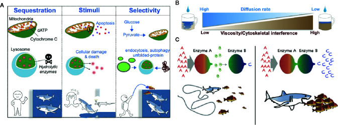
Despite advances in targeted therapeutics and understanding in molecular mechanisms, metastasis remains a substantial obstacle for cancer treatment. Acquired genetic mutations and transcriptional changes can promote the spread of primary tumor cells to distant tissues. Additionally, recent studies have uncovered that metabolic reprogramming of cancer cells is tightly associated with cancer metastasis. However, whether intracellular metabolism is spatially and temporally regulated for cancer cell migration and invasion is understudied. In this review, we highlight the emergence of a concept, termed "membraneless metabolic compartmentalization," as one of the critical mechanisms that determines the metastatic capacity of cancer cells. In particular, we focus on the compartmentalization of purine nucleotide metabolism (e.g., ATP and GTP) at the leading edge of migrating cancer cells through the uniquely phase-separated microdomains where dynamic exchange of nucleotide metabolic enzymes takes place. We will discuss how future insights may usher in a novel class of therapeutics specifically targeting the metabolic compartmentalization that drives tumor metastasis.
Keywords: GTP-metabolism; cancer; leading edge; liquid-liquid phase separation; membraneless metabolic compartmentalization; metabolon; metastasis; purine biosynthesis.
Copyright © 2020 Wolfe, Kamata, Coutinho, Inoue and Sasaki.
Figures

Similar articles
-
Dynamic compartmentalization of purine nucleotide metabolic enzymes at leading edge in highly motile renal cell carcinoma.Biochem Biophys Res Commun. 2019 Aug 13;516(1):50-56. doi: 10.1016/j.bbrc.2019.05.190. Epub 2019 Jun 10. Biochem Biophys Res Commun. 2019. PMID: 31196624 Free PMC article.
-
Membraneless Compartmentalization Facilitates Enzymatic Cascade Reactions and Reduces Substrate Inhibition.ACS Appl Mater Interfaces. 2018 Sep 26;10(38):32782-32791. doi: 10.1021/acsami.8b07573. Epub 2018 Sep 14. ACS Appl Mater Interfaces. 2018. PMID: 30179001 Free PMC article.
-
Phase Separation: Linking Cellular Compartmentalization to Disease.Trends Cell Biol. 2016 Jul;26(7):547-558. doi: 10.1016/j.tcb.2016.03.004. Epub 2016 Apr 1. Trends Cell Biol. 2016. PMID: 27051975 Review.
-
Molecular and Metabolic Reprogramming: Pulling the Strings Toward Tumor Metastasis.Front Oncol. 2021 Jun 3;11:656851. doi: 10.3389/fonc.2021.656851. eCollection 2021. Front Oncol. 2021. PMID: 34150624 Free PMC article. Review.
-
The role of the guanine nucleotide exchange factor Tiam1 in cellular migration, invasion, adhesion and tumor progression.Breast Cancer Res Treat. 2004 Mar;84(1):21-32. doi: 10.1023/B:BREA.0000018421.31632.e6. Breast Cancer Res Treat. 2004. PMID: 14999151 Review.
Cited by
-
Actin cytoskeletal dynamics do not impose an energy drain on growth cone bioenergetics.J Cell Sci. 2023 Aug 15;136(16):jcs261356. doi: 10.1242/jcs.261356. Epub 2023 Aug 18. J Cell Sci. 2023. PMID: 37534394 Free PMC article.
-
Onco-condensates: formation, multi-component organization, and biological functions.Trends Cancer. 2023 Sep;9(9):738-751. doi: 10.1016/j.trecan.2023.05.006. Epub 2023 Jun 20. Trends Cancer. 2023. PMID: 37349246 Free PMC article. Review.
-
Reduced serum high-density lipoprotein cholesterol levels and aberrantly expressed cholesterol metabolism genes in colorectal cancer.World J Clin Cases. 2022 May 16;10(14):4446-4459. doi: 10.12998/wjcc.v10.i14.4446. World J Clin Cases. 2022. PMID: 35663062 Free PMC article.
-
SI-MOIRAI: a new method to identify and quantify the metabolic fate of nucleotides.J Biochem. 2022 Jan 7;170(6):699-711. doi: 10.1093/jb/mvab077. J Biochem. 2022. PMID: 34244779 Free PMC article.
References
Publication types
LinkOut - more resources
Full Text Sources