. 2020 Nov 5:10:557327.
doi: 10.3389/fonc.2020.557327. eCollection 2020.

miR-1260b Activates Wnt Signaling by Targeting Secreted Frizzled-Related Protein 1 to Regulate Taxane Resistance in Lung Adenocarcinoma

Affiliations

miR-1260b Activates Wnt Signaling by Targeting Secreted Frizzled-Related Protein 1 to Regulate Taxane Resistance in Lung Adenocarcinoma

Jin Ren et al. Front Oncol. .

Abstract

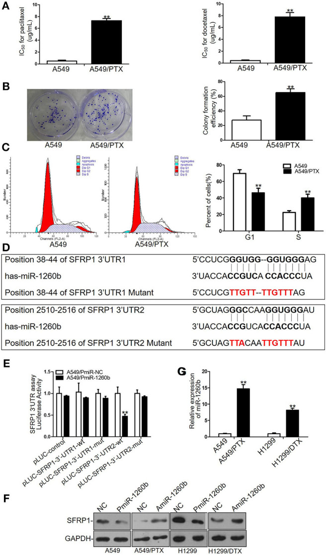
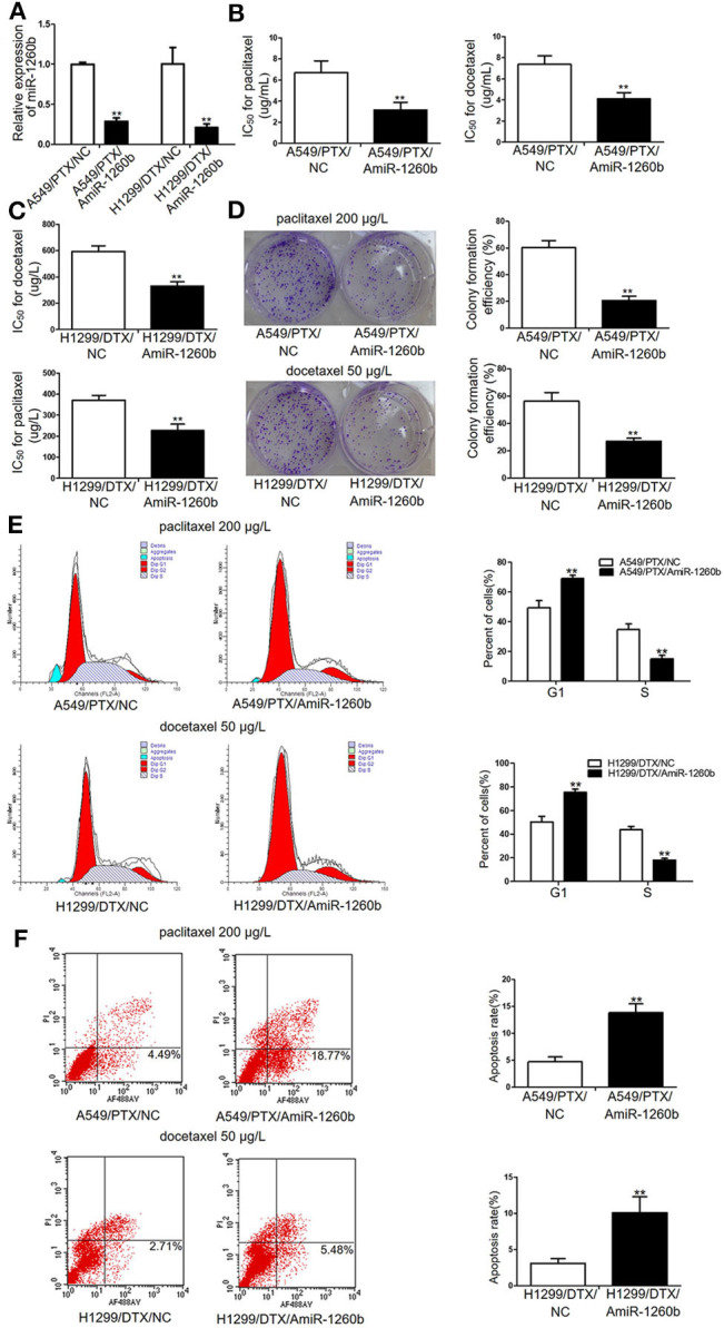
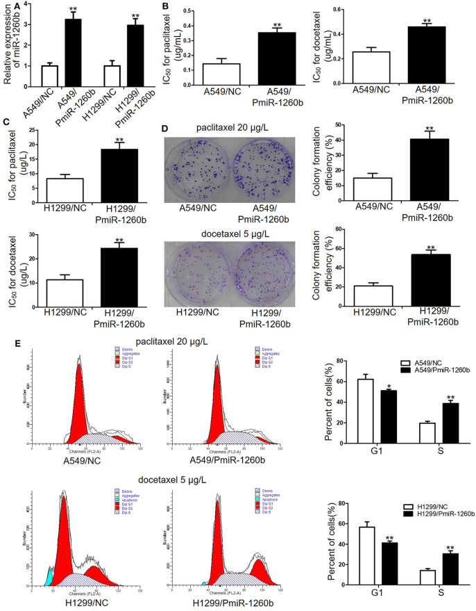
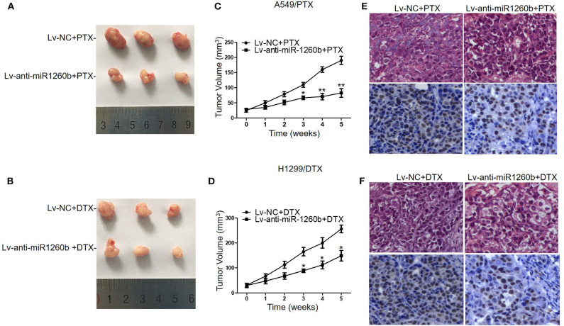
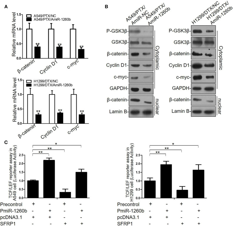
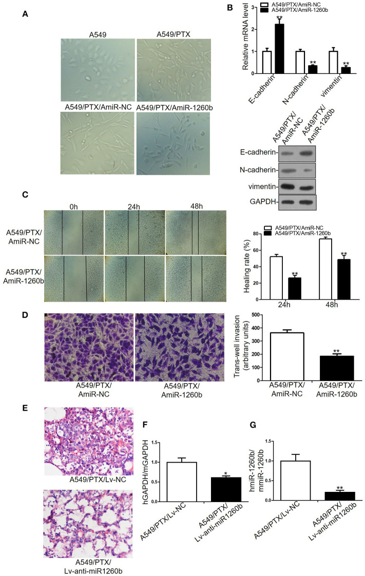
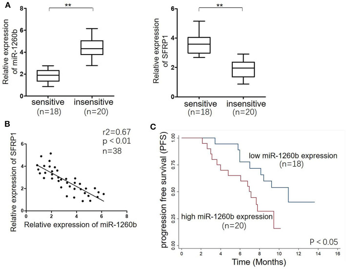
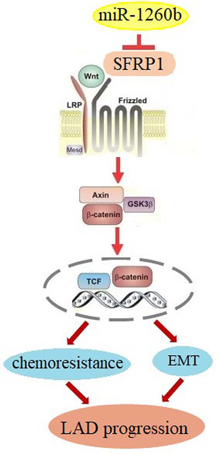
Objectives: MicroRNAs (miRNAs) have been demonstrated to contribute to carcinogenesis; however, their association with tumor chemoresistance is not fully understood. In this study we aimed to investigate the molecular mechanisms involved in resistance to taxane-based chemotherapy in lung adenocarcinoma (LAD). Methods: We established paclitaxel-resistant A549 cells (A549/PTX) and docetaxel-resistant H1299 cells (H1299/DTX). In order to hit the mark, we employed multiple methods including qRT-PCR, western blotting analysis, loss/gain-of-function analysis, luciferase assays, drug sensitivity assays, animal experiment, wound-healing assay, and invasion assay. Results: Bioinformatics analysis and a luciferase reporter assay revealed that secreted frizzled-related protein 1 (SFRP1) is a direct target of miR-1260b. By qRT-PCR analysis, we found that miR-1260b was significantly upregulated in taxane-resistant cells as compared to parental cells. Suppression of miR-1260b reversed the chemoresistance of human LAD cells to taxanes both in vitro and in vivo, whereas ectopic miR-1260b expression decreased the sensitivity of parental LAD cell lines to taxanes. Downregulation of miR-1260b expression inactivated the Wnt signaling pathway and reversed the epithelial-mesenchymal transition (EMT) phenotype of taxane-resistant LAD cells. In clinical tumor tissue samples, high miR-1260b expression was detected in tumors of non-responding patients treated with taxane-based chemotherapy and was associated with low SFRP1 expression and poor prognosis. Conclusions: Our findings reveal that targeting of the miR-1260b/SFRP1/Wnt signaling axis might provide a novel strategy for overcoming chemotherapy resistance in LAD.

Keywords: SFRP1; Wnt signaling; chemoresistance; lung adenocarcinoma; miR-1260; taxane.


Figures

Figure 1
Figure 1
Different sensitivity to paclitaxel and docetaxel between A549/PTX cells and parental A549 cells and SFRP1 was a direct target of miR-1260b in LAD cells. (A) IC50 values for paclitaxel (left) and docetaxel (right) in A549 and A549/PTX cells as determined by MTT assays. (B) Proliferation ability of A549 and A549/PTX cells as determined by colony formation assays. (C) Cell cycle analysis of A549 and A549/PTX cells by flow cytometry. (D) Consensus sequences for miR-1260b in the SFRP1 3′-UTR were predicted by bioinformatics analysis. (E) A reporter vector containing the SFRP1 3′-UTR (wild-type or mutant constructs) was cotransfected with miR-1260b mimic in A549 cells, which were then analyzed using a dual-luciferase reporter assay. (F) SFRP1 protein levels in parental and taxane-resistant cells after transfection with miR-1260b mimic or inhibitor as determined by western blotting. (G) Expression of miR-1260b in A549/PTX and H1299/DTX cells compared with A549 and H1299 cells as indicated by qRT-PCR. miRNA abundance was normalized to the U6 RNA level. The experiment was repeated at least three times. Student t-test was employed to compare the differences between two groups. The data are represented as mean ± SD. **p < 0.01 compared with control group.
Figure 2
Figure 2
Inhibition of miR-1260b reversed the in vitro chemoresistance of A549/PTX and H1299/DTX cells to paclitaxel or docetaxel. (A) Expression levels of miR-1260b in A549/PTX and H1299/DTX cells transfected with mimic NC or miR-1260b inhibitor as determined by qRT-PCR. IC50 values for paclitaxel or docetaxel in A549/PTX (B) and H1299/DTX (C) cells transfected with mimic NC or miR-1260b inhibitor as determined by MTT assays. (D) Colony formation assays revealed proliferation ability of A549/PTX (upper) and H1299/DTX (lower) cells transfected with miR-1260b inhibitor, following treatment of paclitaxel (200 μg/L) or docetaxel (50 μg/L), respectively. Flow cytometric analysis indicated cell cycle (E) and apoptosis (F) in A549/PTX and H1299/DTX cells transfected with mimic NC or miR-1260b inhibitor, following treatment of paclitaxel (200 μg/L) or docetaxel (50 μg/L), respectively. Data between two groups were tested using independent sample t-test. The data are based on 3 to 5 independent experiments and are shown as mean ± SD. **p < 0.01 compared with NC group.
Figure 3
Figure 3
Ectopic miR-1260b expression decreased the chemosensitivity of A549 and H1299 cells to paclitaxel or docetaxel. (A) Expression levels of miR-1260b in A549 and H1299 cells transfected with mimic NC or miR-1260b mimic were determined by qRT-PCR assay. IC50 values for paclitaxel or docetaxel in A549 (B) and H1299 (C) cells transfected with mimic NC or miR-1260b mimic as determined by MTT assays. (D) Colony formation assays revealed proliferation ability of A549 (upper) and H1299 (lower) cells transfected with mimic NC or miR-1260b mimic following treatment of paclitaxel (20 μg/L) or docetaxel (5 μg/L), respectively. (E) Flow cytometry indicated cell cycle of A549 (upper) and H1299 (lower) cells transfected with mimic NC or miR-1260b mimic following treatment of paclitaxel (20 μg/L) or docetaxel (5 μg/L), respectively. Paired t-test was applied to compare the differences between two groups. Similar results were obtained in 3 independent experiments and are shown as mean ± SD. *p < 0.05; **p < 0.01 compared with NC group.
Figure 4
Figure 4
Inhibition of miR-1260b increased the chemosensitivity of A549/PTX and H1299/DTX cells to taxanes in vivo. Nude mice were subcutaneously injected with A549/PTX or H1299/DTX cells that had been infected with lentivirus carrying miR-1260b inhibitor and treated with paclitaxel (15 mg/kg) or docetaxel (1 mg/kg) when the mean tumor volume reached ~100 mm3. (A) Representative picture of tumors from A549/PTX/Lv-NC or A549/PTX/Lv-anti-miR1260b group with paclitaxel treatment. (B) Representative picture of tumors from H1299/DTX/Lv-NC or H1299/DTX/Lv-anti-miR1260b group with docetaxel treatment. (C) Growth curve of tumors derived from A549/PTX/Lv-NC or A549/PTX/Lv-anti-miR1260b cells. (D) Growth curve of tumors derived from H1299/DTX/Lv-NC or H1299/DTX/Lv-anti-miR1260b cells. Paired t-test was performed for comparisons between two groups. Each data point represents the mean ± SD of 7 mice. The three independent experiments gave similar results. *p < 0.05; **p < 0.01 compared with Lv-NC group. (E) H&E- (upper) and PCNA-stained (lower) sections of the transplanted tumors from A549/PTX/Lv-NC or A549/PTX/Lv-anti-miR1260b cells upon paclitaxel treatment. (F) H&E- (upper) and PCNA-stained (lower) sections of the transplanted tumors from H1299/DTX/Lv-NC or H1299/DTX/Lv-anti-miR1260b cells upon docetaxel treatment. Photomicrographs were taken at 400x magnification.
Figure 5
Figure 5
miR-1260b affected Wnt signaling activity by directly targeting SFRP1 in LAD Cells. (A) mRNA expression levels of β-catenin, cyclin D1, and c-myc in A549/PTX and H1299/DTX cells transfected with miR-NC or miR-1260b inhibitor as determined by qRT-PCR. (B) Protein expression levels of p-GSK3β, GSK3β, β-catenin, cyclin D1, and c-myc in A549/PTX and H1299/DTX cells transfected with miR-NC or miR-1260b inhibitor as determined by western blotting. (C) TCF/LEF reporter assay to assess the effect of miR-1260b on Wnt signaling activity in A549 (left) and H1299 (right) cells. Data are represented as mean ± SD of three independent experiments (Student t-test). *p < 0.05; **p < 0.01.
Figure 6
Figure 6
Inhibition of miR-1260b reversed the EMT phenotype and suppressed the invasive ability of A549/PTX cell line. (A) The phenotype of A549, A549/PTX, A549/PTX/AmiR-NC, A549/PTX/AmiR-1260b cells. Photomicrographs were taken at 200x magnification. (B) mRNA and protein expression levels of E-cadherin, N-cadherin, and vimentin in A549/PTX/AmiR-NC and A549/PTX/AmiR-1260b cells as determined by qRT-PCR (left) and western blotting (right). Cell migration and invasion capacities of A549/PTX/AmiR-NC and A549/PTX/AmiR-1260b cells as determined by wound healing (C) and invasion (D) assays, respectively. Photomicrographs were taken at 200x magnification. (E) Eight weeks after tail vein injection of A549/PTX/Lv-NC or A549/PTX/Lv-anti-miR1260b cells, metastatic potential was determined by H&E staining. (F) Human-specific GAPDH (hGAPDH) expression relative to mouse-specific GAPDH (mGAPDH) in circulating tumor cells as determined by qRT-PCR. (G) Human-miR-1260b (hmiR-1260b) expression relative to mouse-miR-1260b (mmiR-1260b) in circulating tumor cells as determined by qRT-PCR. The data are represented as mean ± SD of triplicate samples performed in two independent samples (Student t-test). *p < 0.05; **p < 0.01 compared with control group.
Figure 7
Figure 7
miR-1260b was associated with SFRP1 expression and poor prognosis in LAD tumor tissue samples. (A) mRNA expression levels of miR-1260b (left) and SFRP1 (right) in sensitive (n = 18) and insensitive (n = 20) LAD tissues as detected by qRT-PCR. U6 RNA and GAPDH were used as internal controls. The data express as the mean ± SD (Student t-test). **p < 0.01. (B) Linear regression analysis revealed an inverse correlation between miR-1260b and SFRP1. (C) Kaplan–Meier survival analysis of LAD patients according to the level of miR-1260b expression in tumor tissues. The p-value was determined with the log-rank test.
Figure 8
Figure 8
Schematic diagram for miR-1260b modulation of chemoresistance in NSCLC. miR-1260b regulates chemoresistance by targeting SFRP1 and thereby activating Wnt signaling.

Similar articles

Cited by

  • Fibroblast-derived extracellular vesicles contain SFRP1 and mediate pulmonary fibrosis.
    Burgy O, Mayr CH, Schenesse D, Fousekis Papakonstantinou E, Ballester B, Sengupta A, She Y, Hu Q, Melo-Narvaéz MC, Jain E, Pestoni JC, Mozurak M, Estrada-Bernal A, Onwuka U, Coughlan C, Parimon T, Chen P, Heimerl T, Bange G, Schmeck BT, Lindner M, Hilgendorff A, Ruppert C, Güenther A, Mann M, Yildirim AÖ, Eickelberg O, Jung AL, Schiller HB, Lehmann M, Burgstaller G, Königshoff M. Burgy O, et al. JCI Insight. 2024 Aug 15;9(18):e168889. doi: 10.1172/jci.insight.168889. JCI Insight. 2024. PMID: 39315549 Free PMC article.

References

    1. Siegel RL, Miller KD, Jemal A. Cancer statistics, 2018. CA Cancer J Clin. (2018) 68:7–30. 10.3322/caac.21442 - DOI - PubMed
    1. Goldstraw P, Ball D, Jett JR, Le Chevalier T, Lim E, Nicholson AG, et al. . Non-small-cell lung cancer. Lancet. (2011) 378:1727–40. 10.1016/S0140-6736(10)62101-0 - DOI - PubMed
    1. Zang H, Wang W, Fan S. The role of microRNAs in resistance to targeted treatments of non-small cell lung cancer. Cancer Chemother Pharmacol. (2017) 79:227–31. 10.1007/s00280-016-3130-7 - DOI - PubMed
    1. Joshi M, Liu X, Belani CP. Taxanes, past, present, and future impact on non-small cell lung cancer. Anticancer Drugs. (2014) 25:571–83. 10.1097/CAD.0000000000000080 - DOI - PubMed
    1. Krishnamurthy N, Kurzrock R. Targeting the Wnt/beta-catenin pathway in cancer: update on effectors and inhibitors. Cancer Treat Rev. (2018) 62:50–60. 10.1016/j.ctrv.2017.11.002 - DOI - PMC - PubMed