

Scalable Production of Human Mesenchymal Stromal Cell-Derived Extracellular Vesicles Under Serum-/Xeno-Free Conditions in a Microcarrier-Based Bioreactor Culture System
- PMID: 33224943
- PMCID: PMC7669752
- DOI: 10.3389/fcell.2020.553444
Scalable Production of Human Mesenchymal Stromal Cell-Derived Extracellular Vesicles Under Serum-/Xeno-Free Conditions in a Microcarrier-Based Bioreactor Culture System
Abstract
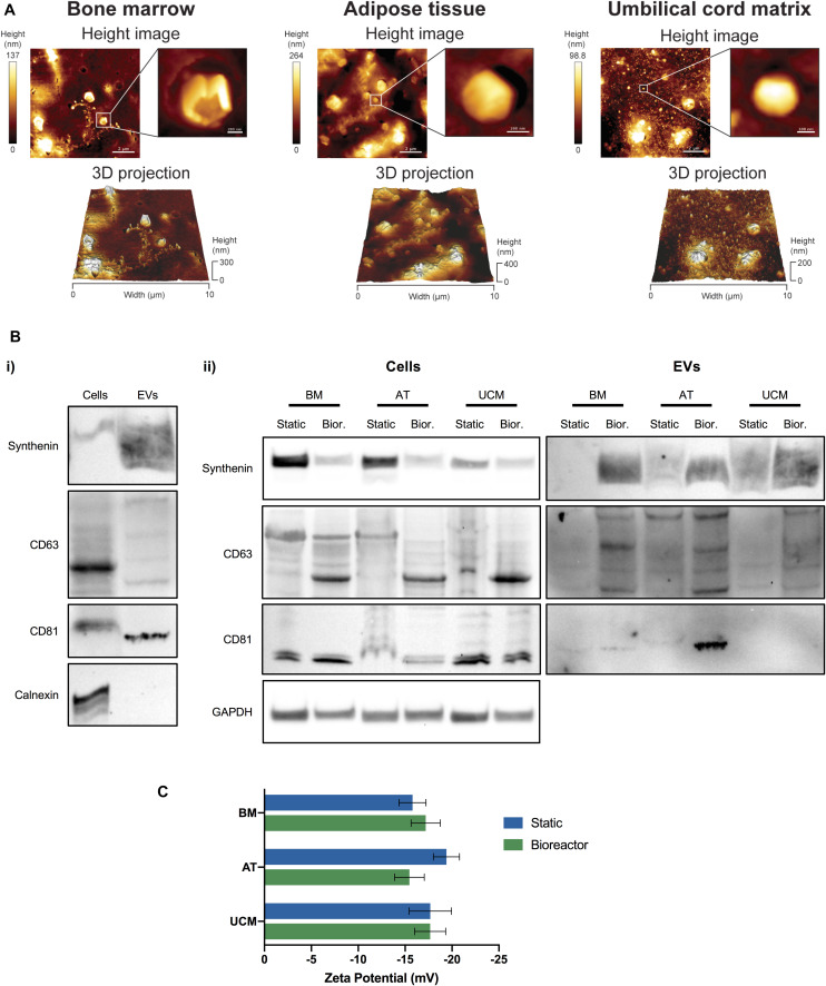
Mesenchymal stromal cells (MSC) hold great promise for tissue engineering and cell-based therapies due to their multilineage differentiation potential and intrinsic immunomodulatory and trophic activities. Over the past years, increasing evidence has proposed extracellular vesicles (EVs) as mediators of many of the MSC-associated therapeutic features. EVs have emerged as mediators of intercellular communication, being associated with multiple physiological processes, but also in the pathogenesis of several diseases. EVs are derived from cell membranes, allowing high biocompatibility to target cells, while their small size makes them ideal candidates to cross biological barriers. Despite the promising potential of EVs for therapeutic applications, robust manufacturing processes that would increase the consistency and scalability of EV production are still lacking. In this work, EVs were produced by MSC isolated from different human tissue sources [bone marrow (BM), adipose tissue (AT), and umbilical cord matrix (UCM)]. A serum-/xeno-free microcarrier-based culture system was implemented in a Vertical-WheelTM bioreactor (VWBR), employing a human platelet lysate culture supplement (UltraGROTM-PURE), toward the scalable production of MSC-derived EVs (MSC-EVs). The morphology and structure of the manufactured EVs were assessed by atomic force microscopy, while EV protein markers were successfully identified in EVs by Western blot, and EV surface charge was maintained relatively constant (between -15.5 ± 1.6 mV and -19.4 ± 1.4 mV), as determined by zeta potential measurements. When compared to traditional culture systems under static conditions (T-flasks), the VWBR system allowed the production of EVs at higher concentration (i.e., EV concentration in the conditioned medium) (5.7-fold increase overall) and productivity (i.e., amount of EVs generated per cell) (3-fold increase overall). BM, AT and UCM MSC cultured in the VWBR system yielded an average of 2.8 ± 0.1 × 10¹¹, 3.1 ± 1.3 × 10¹¹, and 4.1 ± 1.7 × 10¹¹ EV particles (n = 3), respectively, in a 60 mL final volume. This bioreactor system also allowed to obtain a more robust MSC-EV production, regarding their purity, compared to static culture. Overall, we demonstrate that this scalable culture system can robustly manufacture EVs from MSC derived from different tissue sources, toward the development of novel therapeutic products.
Keywords: bioreactors; extracellular vesicles; mesenchymal stromal cells (MSC); scalable production; serum-/xenogeneic-free.
Copyright © 2020 de Almeida Fuzeta, Bernardes, Oliveira, Costa, Fernandes-Platzgummer, Farinha, Rodrigues, Jung, Tseng, Milligan, Lee, Castanho, Gaspar, Cabral and da Silva.
Figures


Similar articles
-
Continuous collection of human mesenchymal-stromal-cell-derived extracellular vesicles from a stirred tank reactor operated under xenogeneic-free conditions for therapeutic applications.Stem Cell Res Ther. 2025 Apr 24;16(1):210. doi: 10.1186/s13287-025-04341-2. Stem Cell Res Ther. 2025. PMID: 40275409 Free PMC article.
-
Dual production of human mesenchymal stromal cells and derived extracellular vesicles in a dissolvable microcarrier-based stirred culture system.Cytotherapy. 2024 Jul;26(7):749-756. doi: 10.1016/j.jcyt.2024.03.001. Epub 2024 Mar 11. Cytotherapy. 2024. PMID: 38506771
-
Integrated culture platform based on a human platelet lysate supplement for the isolation and scalable manufacturing of umbilical cord matrix-derived mesenchymal stem/stromal cells.J Tissue Eng Regen Med. 2017 May;11(5):1630-1640. doi: 10.1002/term.2200. Epub 2016 Jul 22. J Tissue Eng Regen Med. 2017. PMID: 27444977
-
From Mesenchymal Stromal Cells to Engineered Extracellular Vesicles: A New Therapeutic Paradigm.Front Cell Dev Biol. 2021 Jul 20;9:705676. doi: 10.3389/fcell.2021.705676. eCollection 2021. Front Cell Dev Biol. 2021. PMID: 34409037 Free PMC article. Review.
-
Isolation, cultivation, and characterization of human mesenchymal stem cells.Cytometry A. 2018 Jan;93(1):19-31. doi: 10.1002/cyto.a.23242. Epub 2017 Oct 26. Cytometry A. 2018. PMID: 29072818 Review.
Cited by
-
Advances in the Field of Micro- and Nanotechnologies Applied to Extracellular Vesicle Research: Take-Home Message from ISEV2021.Micromachines (Basel). 2021 Dec 16;12(12):1563. doi: 10.3390/mi12121563. Micromachines (Basel). 2021. PMID: 34945413 Free PMC article.
-
Characterization of the Aeration and Hydrodynamics in Vertical-Wheel™ Bioreactors.Bioengineering (Basel). 2022 Aug 12;9(8):386. doi: 10.3390/bioengineering9080386. Bioengineering (Basel). 2022. PMID: 36004911 Free PMC article.
-
Neurodifferentiation and Neuroprotection Potential of Mesenchymal Stromal Cell-Derived Secretome Produced in Different Dynamic Systems.Biomedicines. 2023 Apr 22;11(5):1240. doi: 10.3390/biomedicines11051240. Biomedicines. 2023. PMID: 37238911 Free PMC article.
-
Equine mesenchymal stem cell-derived extracellular vesicle productivity but not overall yield is improved via 3-D culture with chemically defined media.J Am Vet Med Assoc. 2024 Apr 1;262(S1):S97-S108. doi: 10.2460/javma.24.01.0001. Print 2024 Jun 1. J Am Vet Med Assoc. 2024. PMID: 38547591 Free PMC article.
-
vCPP2319 interacts with metastatic breast cancer extracellular vesicles (EVs) and transposes a human blood-brain barrier model.Heliyon. 2024 Dec 4;10(23):e40907. doi: 10.1016/j.heliyon.2024.e40907. eCollection 2024 Dec 15. Heliyon. 2024. PMID: 39717586 Free PMC article.
References
LinkOut - more resources
Full Text Sources
Other Literature Sources
Miscellaneous