

Enhanced bodily states of fear facilitates bias perception of fearful faces
- PMID: 33225980
- PMCID: PMC7682010
- DOI: 10.1186/s13041-020-00674-6
Enhanced bodily states of fear facilitates bias perception of fearful faces
Abstract
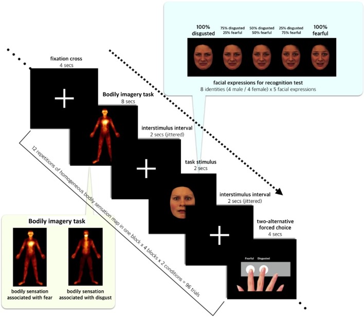
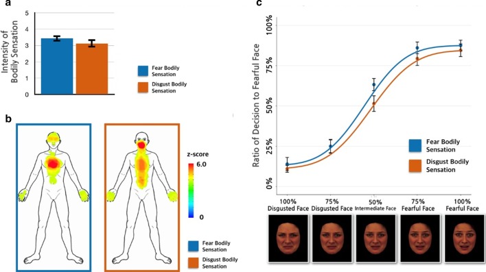
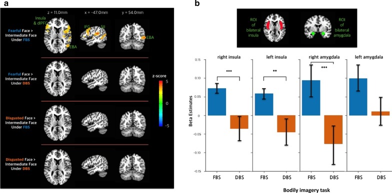
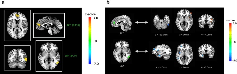
We investigated whether enhanced interoceptive bodily states of fear would facilitate recognition of the fearful faces. Participants performed an emotional judgment task after a bodily imagery task inside a functional magnetic resonance imaging scanner. In the bodily imagery task, participants were instructed to imagine feeling the bodily sensations of two specific somatotopic patterns: a fear-associated bodily sensation (FBS) or a disgust-associated bodily sensation (DBS). They were shown faces expressing various levels of fearfulness and disgust and instructed to classify the facial expression as fear or disgust. We found a stronger bias favoring the "fearful face" under the congruent FBS condition than under the incongruent DBS condition. The brain response to fearful versus intermediate faces increased in the fronto-insular-temporal network under the FBS condition, but not the DBS condition. The fearful face elicited activity in the anterior cingulate cortex and extrastriate body area under the FBS condition relative to the DBS condition. Furthermore, functional connectivity between the anterior cingulate cortex/extrastriate body area and the fronto-insular-temporal network was modulated according to the specific bodily sensation. Our findings suggest that somatotopic patterns of bodily sensation provide informative access to the collective visceral state in the fear processing via the fronto-insular-temporal network.
Keywords: Anterior cingulate cortex; Emotional face; Extrastriate body area; Fear; Interoception.
Conflict of interest statement
The authors declare that they have no competing interests.
Figures
Similar articles
-
Enhanced Expectation of External Sensations of the Chest Regulates the Emotional Perception of Fearful Faces.Brain Sci. 2021 Jul 19;11(7):946. doi: 10.3390/brainsci11070946. Brain Sci. 2021. PMID: 34356181 Free PMC article.
-
Bodily Information and Top-Down Affective Priming Jointly Affect the Processing of Fearful Faces.Front Psychol. 2021 Jun 2;12:625986. doi: 10.3389/fpsyg.2021.625986. eCollection 2021. Front Psychol. 2021. PMID: 34149514 Free PMC article.
-
Differential neural responses to overt and covert presentations of facial expressions of fear and disgust.Neuroimage. 2004 Apr;21(4):1484-96. doi: 10.1016/j.neuroimage.2003.12.013. Neuroimage. 2004. PMID: 15050573
-
Distributed and interactive brain mechanisms during emotion face perception: evidence from functional neuroimaging.Neuropsychologia. 2007 Jan 7;45(1):174-94. doi: 10.1016/j.neuropsychologia.2006.06.003. Epub 2006 Jul 18. Neuropsychologia. 2007. PMID: 16854439 Review.
-
Cellular activity in insular cortex across seconds to hours: Sensations and predictions of bodily states.Neuron. 2021 Nov 17;109(22):3576-3593. doi: 10.1016/j.neuron.2021.08.036. Epub 2021 Sep 27. Neuron. 2021. PMID: 34582784 Free PMC article. Review.
Cited by
-
Impaired cardiac modulation in patients with functional seizures: Results from a face intensity judgment task.Epilepsia. 2023 Nov;64(11):3073-3081. doi: 10.1111/epi.17761. Epub 2023 Sep 12. Epilepsia. 2023. PMID: 37611952 Free PMC article.
-
Enhanced Expectation of External Sensations of the Chest Regulates the Emotional Perception of Fearful Faces.Brain Sci. 2021 Jul 19;11(7):946. doi: 10.3390/brainsci11070946. Brain Sci. 2021. PMID: 34356181 Free PMC article.
-
General and anxiety-linked influences of acute serotonin reuptake inhibition on neural responses associated with attended visceral sensation.Transl Psychiatry. 2024 Jun 6;14(1):241. doi: 10.1038/s41398-024-02971-3. Transl Psychiatry. 2024. PMID: 38844469 Free PMC article. Clinical Trial.
References
-
- James W: What is an emotion? Mind 1884, os-IX(34):188–205.
Publication types
MeSH terms
LinkOut - more resources
Full Text Sources
