. 2021 Jan;27(1):134-146.
doi: 10.5152/dir.2020.19515.

MRI in patients with urethral stricture: a systematic review

Affiliations

MRI in patients with urethral stricture: a systematic review

Mikolaj Frankiewicz et al. Diagn Interv Radiol. 2021 Jan.

Abstract

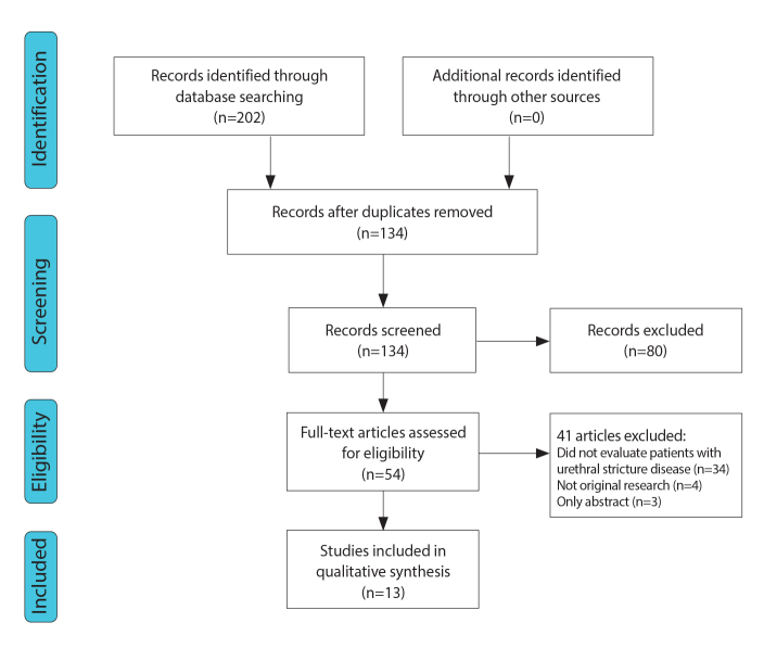
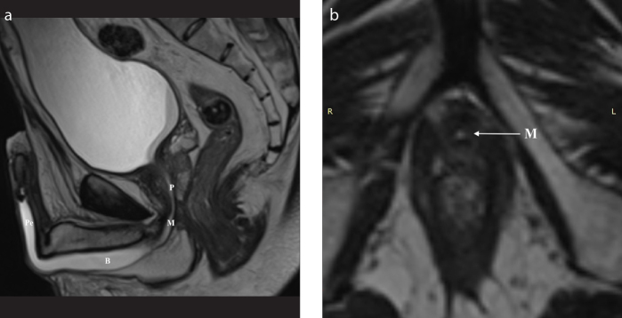
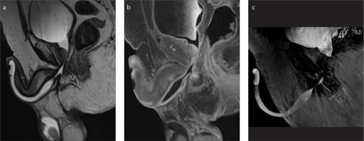
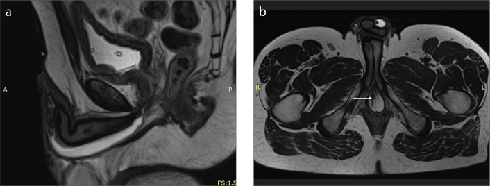
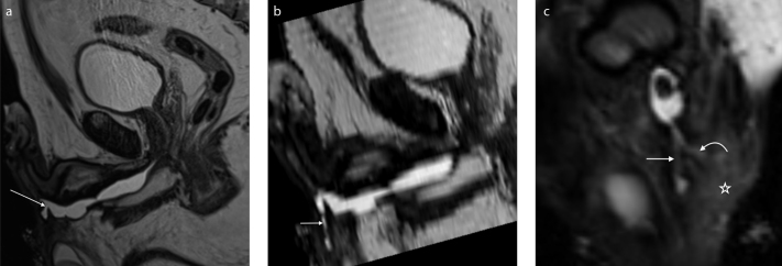
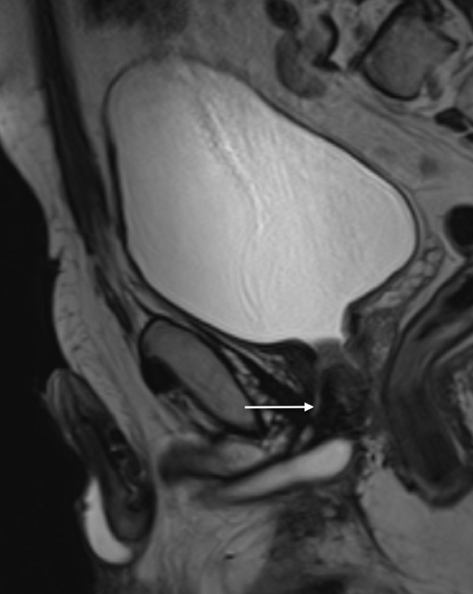
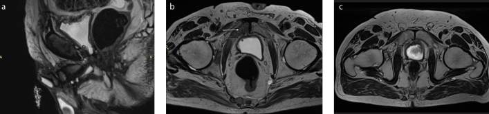
Magnetic resonance imaging (MRI) is gaining acceptance as a diagnostic tool in urethral stricture disease. Numerous publications emphasize on the advantages of MRI including its ability to determine periurethral spongiofibrosis, thus overcoming the main limitation of retrograde urethrography (RUG). It is also becoming an alternative for sonourethrography (SUG), which is a highly subjective examination. Magnetic resonance urethrography (MRU) has become an increasingly appreciated tool for diagnosing patients with urethral stricture disease. Obtained data provides radiologists and urethral reconstructive surgeons with additional information regarding anatomical relationships and periurethral tissue details, facilitating further treatment planning. Considering the great prevalence of urethral stricture disease and necessity of using accurate, and acceptable diagnostic method, this review was designed to provide radiologists and clinicians with a systematic review of the literature on the use of MRI in the urethral stricture disease.


Conflict of interest statement

Conflict of interest disclosure

The authors declared no conflicts of interest.

Figures

Figure 1
Figure 1
PRISMA flowchart.
Figure 2. a, b
Figure 2. a, b
Imaging anatomy of the male urethra in a 35-year-old man. T2-weighted sagittal image (a) after distention of the urethra with sterile gel shows the prostatic urethra (P), level of the membranous urethra (M), and both bulbar (B) and penile (P) parts of the anterior urethra. T2-weighted axial image (b) at the level of the genitourinary membrane shows urethra as a small round structure of high signal intensity (arrow).
Figure 3. a–c
Figure 3. a–c
A 72-year-old patient who developed an urethral stricture due to straddle injury followed by multiple interventions, currently after removal of chronic Foley catheter. Status post transurethral resection of the prostate (TURP). T2-weighted sagittal image (a) shows a 24.5 mm stricture of the bulbar urethra—the site and extension of the stenosis are well depicted by the distended urethra. After intravenous administration of contrast agent, the activity of the inflammatory process can be evaluated (b). MIP reconstruction is also enclosed (c).
Figure 4. a, b
Figure 4. a, b
T2-weighted (a) and contrast-enhanced T1-weighted (b) sagittal images show long stenosis of the bulbar and penile segments in a 39-year-old patient.
Figure 5. a, b
Figure 5. a, b
A 35-year-old patient with post-traumatic urethral stenosis. T2-weighted sagittal image (a) shows severe stenosis of the bulbar urethra and axial image (b) shows a fistula in the right penile bulb (arrow). The fistula was limited to the right penile bulb.
Figure 6. a–c
Figure 6. a–c
A 59-year-old patient with multiple stenoses of the urethra complicated by a branching urethrocutaneous fistula. T2-weighted sagittal image (a) shows the origin of the urethrocutaneous fistula (arrow). Parasagittal reformatted image (b) shows the main tract of the fistula (short arrow). Paracoronal reformatted image (c) shows a second branch of the fistula (curved arrow). Signs of inflammation of the adjacent soft tissue are also visible (asterisk).
Figure 7
Figure 7
T2-weighted sagittal image shows presence of a false track (arrow) in a 59-year-old patient referred to our institution with suspicion of urethral stenosis.
Figure 8. a–c
Figure 8. a–c
A 74-year-old patient who underwent radical prostatectomy was examined with MRI to evaluate urethral stricture associated with radiation therapy. T2-weighted sagittal (a) and axial (b) images show focal lesions within the minor pelvis bones (arrow) and lumbar spine suspected of being metastases, not present in a previous examination 10 months earlier as shown in T2-weighted axial image (c).

Similar articles

Cited by

References

    1. Jordan GH, Schlossberg SM. Surgery of the penis and urethra. In: Wein AJ, et al., editors. Campbell-Walsh Urology. 9th ed. Vol. 1. Philadelphia, Pa: WB Saunders Co; 2007. pp. 1023–1097.
    1. Mundy AR. Management of urethral strictures. Postgrad Med J. 2006;82:489–499. doi: 10.1136/pgmj.2005.042945. - DOI - PMC - PubMed
    1. Alwaal A, Blaschko SD, McAninch JW, Breyer BN. Epidemiology of urethral strictures. Transl Androl Urol. 2014;3:209–213. - PMC - PubMed
    1. Baskin LS, Constantinescu SC, Howard PS. Biochemical characterization and quantitation of the collagenous components of urethral stricture tissue. J Urol. 1993;150:426–647. doi: 10.1016/S0022-5347(17)35572-6. - DOI - PubMed
    1. Wood DN, Andrich DE, Greenwell TJ, Mundy AR. Standing the test of time: The long-term results of urethroplasty. World J Urol. 2006;24:250–254. doi: 10.1007/s00345-006-0057-3. - DOI - PubMed

Publication types

LinkOut - more resources