Successful management of gastric remnant necrosis after proximal gastrectomy using a double elementary diet tube: a case report
- PMID: 33226508
- PMCID: PMC7683626
- DOI: 10.1186/s40792-020-01056-9
Successful management of gastric remnant necrosis after proximal gastrectomy using a double elementary diet tube: a case report
Abstract
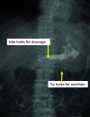
Background: The stomach has many incoming vessels and is resistant to ischemia due to the rich microvascular network within its submucosal layer. Although reports of gastric remnant necrosis after gastrectomy have been rare, mortality rates remain substantially high when present. A double elementary diet (W-ED) tube, which can be used for both enteral feeding and gastrointestinal tract decompression, has been developed for anastomotic leakage and postoperative nutritional management after upper gastrointestinal surgery. The current report presents a case of gastric remnant necrosis after proximal gastrectomy that was successfully managed through conservative treatment with a W-ED tube.
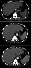
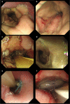
Case presentation: A 73-year-old male was referred to our hospital for an additional resection after endoscopic submucosal dissection (ESD) for gastric cancer. Endoscopic findings showed an ESD scar on the posterior wall of the upper portion of the stomach, while computed tomography (CT) showed no obvious regional lymph node enlargement and distant metastases. The patient subsequently underwent laparoscopic proximal gastrectomy and esophagogastrostomy but developed candidemia on postoperative day 7. On postoperative day 14, endoscopy revealed gastric ischemic changes around the anastomotic site, suggesting that the patient's candidemia developed due to gastric necrosis. His vital signs remained normal, while the gastric remnant ischemia was localized. Given that surgery in the presence of candidemia was considered extremely risky, conservative treatment was elected. A W-ED tube was placed nasally, after which enteral feeding was initiated along with gastrointestinal tract decompression. Although the patient subsequently developed anastomotic leakage due to gastric remnant necrosis, local control was achieved and conservative treatment was continued. On postoperative day 52, healing of the gastric remnant necrosis and anastomotic leakage was confirmed, after which the patient started drinking water. Although balloon dilation was required due to anastomotic stenosis, the patient was able to resume oral intake and was discharged on postoperative day 88.
Conclusions: Herein, we present our experience with a case of gastric remnant necrosis after proximal gastrectomy, wherein conservative management was achieved using a W-ED tube. In cases involving high operative risk, the management should be mindful of gastric remnant necrosis as a post-gastrectomy complication.
Keywords: Conservative treatment; Double elementary diet tube; Gastric remnant necrosis; Proximal gastrectomy.
Conflict of interest statement
The authors declare that they have no competing interests.
Figures


References
LinkOut - more resources
Full Text Sources
Miscellaneous

