Review
. 2021 Jan 15:891:173748.
doi: 10.1016/j.ejphar.2020.173748. Epub 2020 Nov 20.

A review on possible mechanistic insights of Nitazoxanide for repurposing in COVID-19

Affiliations
Review

A review on possible mechanistic insights of Nitazoxanide for repurposing in COVID-19

Amit S Lokhande et al. Eur J Pharmacol. .

Abstract

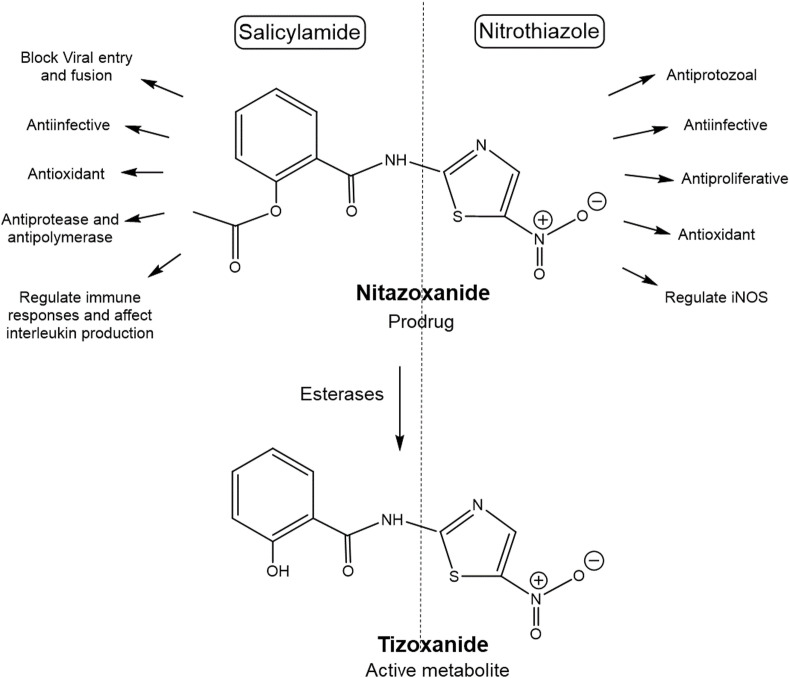
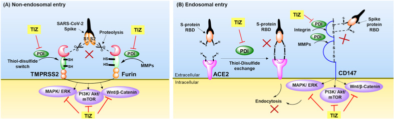
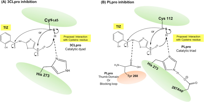
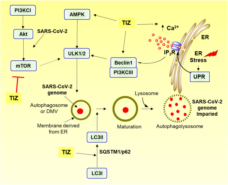
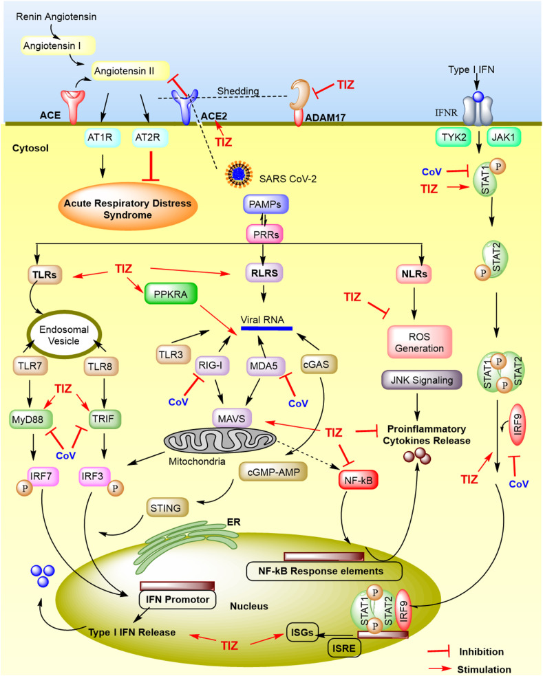
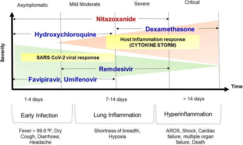
The global pandemic of Coronavirus Disease 2019 (COVID-19) has brought the world to a grinding halt. A major cause of concern is the respiratory distress associated mortality attributed to the cytokine storm. Despite myriad rapidly approved clinical trials with repurposed drugs, and time needed to develop a vaccine, accelerated search for repurposed therapeutics is still ongoing. In this review, we present Nitazoxanide a US-FDA approved antiprotozoal drug, as one such promising candidate. Nitazoxanide which is reported to exert broad-spectrum antiviral activity against various viral infections, revealed good in vitro activity against SARS-CoV-2 in cell culture assays, suggesting potential for repurposing in COVID-19. Furthermore, nitazoxanide displays the potential to boost host innate immune responses and thereby tackle the life-threatening cytokine storm. Possibilities of improving lung, as well as multiple organ damage and providing value addition to COVID-19 patients with comorbidities, are other important facets of the drug. The review juxtaposes the role of nitazoxanide in fighting COVID-19 pathogenesis at multiple levels highlighting the great promise the drug exhibits. The in silico data and in vitro efficacy in cell lines confirms the promise of nitazoxanide. Several approved clinical trials world over further substantiate leveraging nitazoxanide for COVID-19 therapy.

Keywords: Antiviral; COVID-19; Clinical trials; Immunomodulation; Nitazoxanide; SARS-CoV-2.


Figures

Fig. 1
Fig. 1
Nitazoxanide structural features. Nitazoxanide a nitrothiazolyl benzamide is a prodrug metabolized to deacetylated active tizoxanide (TIZ). Both have the two major structural components a) salicylamide moiety and b) nitrothiazole moiety which are responsible for the multiple therapeutic actions. iNOS, instrinsic nitric oxide synthase.
Fig. 2
Fig. 2
Schematic representation of possible role of tizoxanide (TIZ)-inhibition of SARS-CoV-2 fusion and entry. (A) Non-endosomal entry involves the proteolytic cleavage of SARS-CoV-2 spike proteins (S1/S2) by host cell proteases TMPRSS2 and furin. TIZ inhibits PDI and affects thiol-disulfide oxidoreductase switch in the ectodomains of TMPRSS2, and inhibition of PDI also inhibits MMPs activation required for furin action, (B) Endosomal entry involves the recognition and binding of RBD of SARS-CoV-2 spike proteins to the host cell receptors ACE2 and CD147 enabling receptor mediated endocytic entry. TIZ inhibits thiol-disulfide exchange mechanism essential for receptor interaction with RBD by inhibiting PDI. This PDI inhibition also hampers MMPs and intergrins activation required for CD-147 activity. TIZ also inhibits intracellular signalling MAPK/ERK, PI3K/Akt/mTOR and Wnt/β-catenin affecting SARS-CoV-2 fusion and entry. ACE2, Angiotensin converting enzyme 2; CD147, Cluster of differentiation 147; MAPK/ERK, Mitogen-activated protein kinase/extracellular signal-regulated kinase; MMPs, Matrix metalloproteinases; PDI, Protein disulfide isomerase; PI3K/Akt/mTOR, phosphatidylinositol-3-kinase/protein kinase B/mammalian target of rapamycin; RBD, Receptor binding domain; TIZ, Tizoxanide; TMPRSS2, Transmembrane protease serine 2.
Fig. 3
Fig. 3
Schematic representation of nitazoxanide antiprotease action against 3CLpro and PLpro of SARS-CoV-2. Tizoxanide (TIZ) (active metabolite of nitazoxanide) interacts with cysteine residue and thus inhibits the enzyme activity, (A) 3CLpro inhibition and (B) PLpro inhibition. Nucleophilic cysteine thiol interacts with electrophilic moieties of tizoxanide (TIZ). 3CLpro, 3-chymotrypsin like protease; PLpro, Papain like protease.
Fig. 4
Fig. 4
Schematic representation of tizoxanide induced autophagy hindering SARS-CoV-2 genome synthesis. Tizoxanide (TIZ) activates Beclin1, PI3KCIII and AMPK which leads to activation of ULK1 inducing autophagosome formation. TIZ also increases cytosolic Ca2+ concentration through IP3R by depleting ER Ca2+ store inducing ER stress and activating protective UPR signalling. TIZ causes conversion of LC3I to LC3II, degradation of p62/SQSTM1 and inhibits PI3KCI/Akt/mTOR signalling, all together promoting autophagy. AMPK, AMP-protein activated kinase; ER, Endoplasmic reticulum; IP3R, Inositol trisphosphate receptor; LC3, Light chain 3; p62/SQSTM1, cargo receptor p62/sequestome1; PI3KCI, Phosphatidylinositol-3-kinase class I; PI3KCIII, Phosphatidylinositol-3-kinase class III; PI3KCI/Akt/mTOR, phosphatidylinositol-3-kinase/protein kinase B/mammalian target of rapamycin; ULK1/2, Unc-51-like kinase-1/2; UPR, Unfolded protein response.
Fig. 5
Fig. 5
Evasion of host innate immune responses by SARS-CoV-2 and stimulation of host antiviral immune responses by tizoxanide (TIZ). SARS-CoV-2 entry via ACE2 is hampered by TIZ which regulates renin-angiotensin system by blocking angiotensin II binding to its receptors (AT1R and AT2R), to provide protective responses. ADAM-17 mediated ACE2 shedding is prevented by TIZ due to PDI inhibition, thus curbing lung inflammation and consequently ARDS. Upon entry, SARS-CoV-2 pathogen-associated molecular patterns (PAMPs) evades recognition by host cells pattern-recognition-receptors (PRRs), like RLRs, TLRs and NLRs, which is opposed by TIZ. These PRRs cause downstream activation of NF-κB, IRF3/7 signalling, which leads to the secretion of proinflammatory cytokines and type I-IFN respectively. Secreted IFN binds to IFN receptor activating JAK/STAT signalling, which forms a complex with IRF9 and produces interferon stimulated genes (ISGs) promoted under IFN-stimulated response element (ISRE), boosting antiviral innate immune responses, enabling virus clearance. TIZ activates Type-I IFN secretion and curb proinflammatory responses. ACE2, Angiotensin converting enzyme 2; ADAM-17, A disintegrin and metallopeptidase domain 17; ARDS, Acute respiratory distress syndrome; AT1R, Angiotensin II receptor type 1; AT2R, Angiotensin II receptor type 2; cGAS, cyclic GMP-AMP synthase; IFN, Interferon; IFNR, IFNα/β receptor; IRF3/7, Interferon regulatory factor 3/7; JAK, Janus kinase; MAVS, Mitochondrial antiviral-signalling protein; MDA5, Melanoma differentiation-associated gene 5; NF-κB, Nuclear factor-κB; NLRs, Nucleotide-binding oligomerization domain-(NOD)-like receptors; PDI, Protein disulfide isomerase; PPKRA, Phopsphorylated protein kinase receptor; RLRs, Retinoic acid-inducible gene I protein (RIG-I) like receptors; ROS, Reactive oxygen species; STAT, Signal transducer and activator of transcription; STING, stimulator of interferon genes protein; TLRs, Toll-like receptors; TRIF, TIR-domain-containing adaptor protein including IFN-β; TYK2, Tyrosine kinase 2.
Fig. 6
Fig. 6
Different clinical phases of COVID-19 infection based on severity and recommended drug use. ARDS, Acute respiratory distress syndrome.

References

    1. Abdul Kadhim A.H., Hadi N.R., Abdulhussein M. Preprocessing of the candidate antiviral drugs against COVID-19 in models of SARS cov2 targets. La Prensa Medica Argentina. 2020;106:2. doi: 10.1002/ddr.21701. - DOI
    1. Aftab S.O., Ghouri M.Z., Masood M.U., Haider Z., Khan Z., Ahmad A., Munawar N. Analysis of SARS-CoV-2 RNA-dependent RNA polymerase as a potential therapeutic drug target using a computational approach. J. Transl. Med. 2020;18:275. doi: 10.1186/s12967-020-02439-0. - DOI - PMC - PubMed
    1. Agajanian M.J., Walker M.P., Axtman A.D., Ruela-de-Sousa R.R., Serafin D.S., Rabinowitz A.D., Graham D.M., Ryan M.B., Tamir T., Nakamichi Y., Gammons M.V., Bennett J.M., Couñago R.M., Drewry D.H., Elkins J.M., Gileadi C., Gileadi O., Godoi P.H., Kapadia N., Müller S., Santiago A.S., Sorrell F.J., Wells C.I., Fedorov O., Willson T.M., Zuercher W.J., Major M.B. WNT activates the AAK1 kinase to promote clathrin-mediated endocytosis of LRP6 and establish a negative feedback loop. Cell Rep. 2019;26:79–93. doi: 10.1016/j.celrep.2018.12.023. e8. - DOI - PMC - PubMed
    1. Agostini M.L., Andres E.L., Sims A.C., Graham R.L., Sheahan T.P., Lu X., Smith E.C., Case J.B., Feng J.Y., Jordan R., Ray A.S., Cihlar T., Siegel D., Mackman R.L., Clarke M.O., Baric R.S., Denison M.R. Coronavirus susceptibility to the antiviral remdesivir (GS-5734) is mediated by the viral polymerase and the proofreading exoribonuclease. mBio. 2018;9 doi: 10.1128/mBio.00221-18. e00221-18. - DOI - PMC - PubMed
    1. Alharbi A.F., Parrington J. Endolysosomal Ca2+ signaling in cancer: the role of TPC2, from tumorigenesis to metastasis. Front. Cell Dev. Biol. 2019;7:302. doi: 10.3389/fcell.2019.00302. - DOI - PMC - PubMed