

A photo-crosslinkable cartilage-derived extracellular matrix bioink for auricular cartilage tissue engineering
- PMID: 33227486
- PMCID: PMC7855948
- DOI: 10.1016/j.actbio.2020.11.029
A photo-crosslinkable cartilage-derived extracellular matrix bioink for auricular cartilage tissue engineering
Abstract
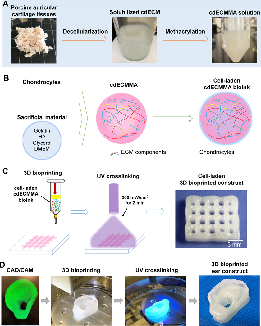
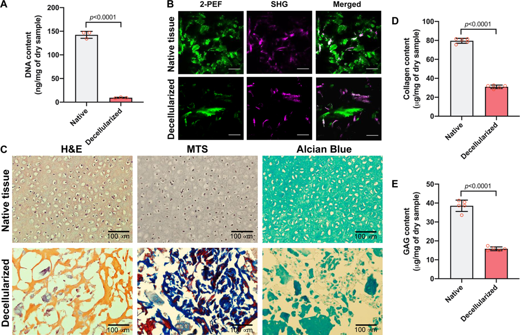
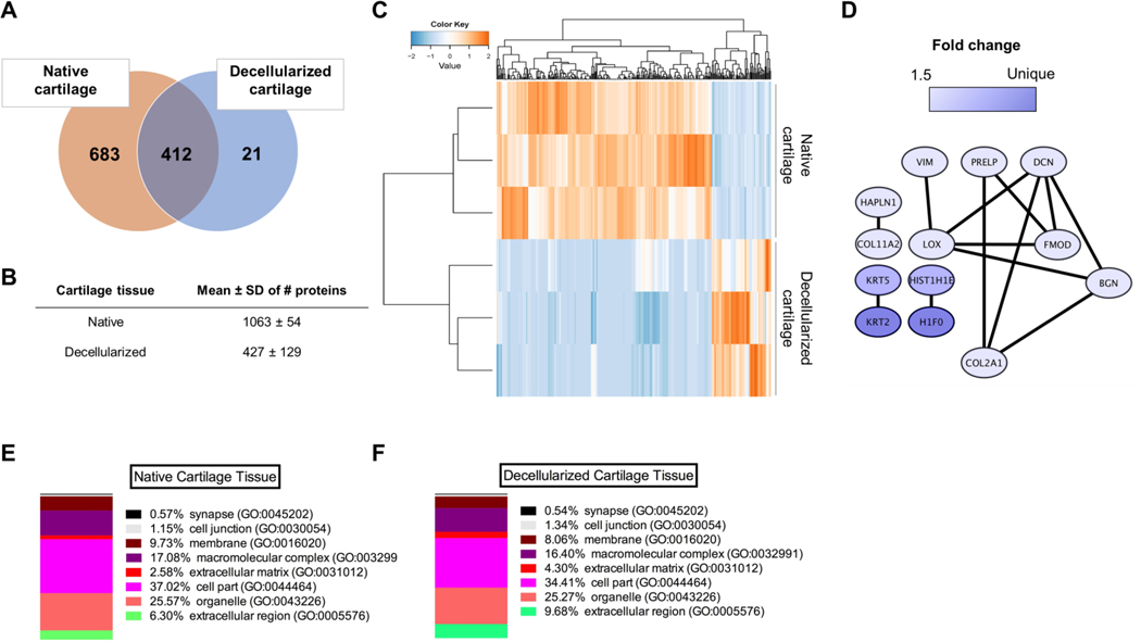
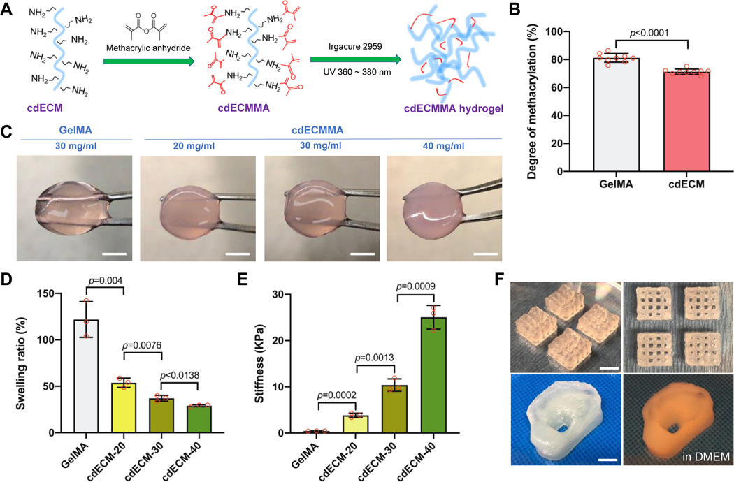
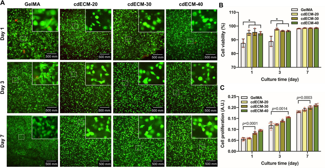
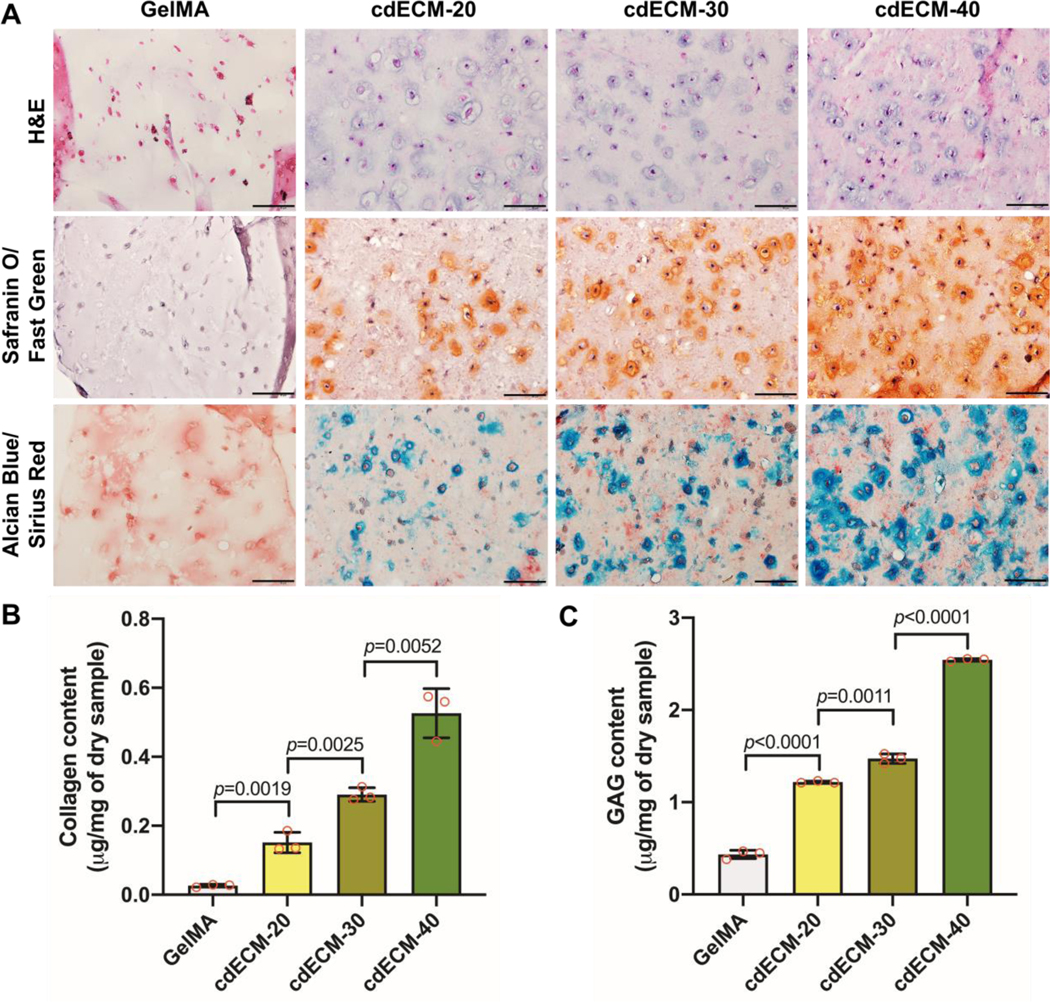
Three-dimensional (3D) bioprinting of patient-specific auricular cartilage constructs could aid in the reconstruction process of traumatically injured or congenitally deformed ear cartilage. To achieve this, a hydrogel-based bioink is required that recapitulates the complex cartilage microenvironment. Tissue-derived decellularized extracellular matrix (dECM)-based hydrogels have been used as bioinks for cell-based 3D bioprinting because they contain tissue-specific ECM components that play a vital role in cell adhesion, growth, and differentiation. In this study, porcine auricular cartilage tissues were isolated and decellularized, and the decellularized cartilage tissues were characterized by histology, biochemical assay, and proteomics. This cartilage-derived dECM (cdECM) was subsequently processed into a photo-crosslinkable hydrogel using methacrylation (cdECMMA) and mixed with chondrocytes to create a printable bioink. The rheological properties, printability, and in vitro biological properties of the cdECMMA bioink were examined. The results showed cdECM was obtained with complete removal of cellular components while preserving major ECM proteins. After methacrylation, the cdECMMA bioinks were printed in anatomical ear shape and exhibited adequate mechanical properties and structural integrity. Specifically, auricular chondrocytes in the printed cdECMMA hydrogel constructs maintained their viability and proliferation capacity and eventually produced cartilage ECM components, including collagen and glycosaminoglycans (GAGs). The potential of cell-based bioprinting using this cartilage-specific dECMMA bioink is demonstrated as an alternative option for auricular cartilage reconstruction.
Keywords: Bioink; Bioprinting; Cartilage tissue engineering; Decellularization; Extracellular matrix; Methacrylation.
Copyright © 2020 Acta Materialia Inc. Published by Elsevier Ltd. All rights reserved.
Conflict of interest statement
Declaration of Competing Interest The authors declare that they have no known competing financial interests or personal relationships that could have appeared to influence the work reported in this paper.
Figures
Comment in
-
Response to Letter to Editor "Comment on 'A photo-crosslinkable cartilage-derived extracellular matrix bioink for auricular cartilage tissue engineering' by Visscher et al.".Acta Biomater. 2021 Nov;135:724. doi: 10.1016/j.actbio.2021.09.059. Epub 2021 Oct 1. Acta Biomater. 2021. PMID: 34601103
-
Comment on "A photo-crosslinkable cartilage-derived extracellular matrix bioink for auricular cartilage tissue engineering" by Visscher et al.Acta Biomater. 2021 Nov;135:723. doi: 10.1016/j.actbio.2021.09.058. Epub 2021 Oct 1. Acta Biomater. 2021. PMID: 34601104
References
-
- Cole A, A review of diversity in the evolution and development of cartilage: the search for the origin of the chondrocyte, Eur Cell Mater 21(122) (2011) 9. - PubMed
-
- Soukup B, Mashhadi SA, Bulstrode NW, Health-related quality-of-life assessment and surgical outcomes for auricular reconstruction using autologous costal cartilage, Plastic and reconstructive surgery 129(3) (2012) 632–640. - PubMed
-
- Kludt NA, Vu H, Auricular reconstruction with prolonged tissue expansion and porous polyethylene implants, Annals of plastic surgery 72 (2014) S14–S17. - PubMed
-
- Fernandes JR, Driscoll DN, Burn ear reconstruction using porous polyethylene implants and tissue expansion, Journal of Burn Care & Research 37(4) (2016) e348–e352. - PubMed
Publication types
MeSH terms
Grants and funding
LinkOut - more resources
Full Text Sources
Other Literature Sources
