Review
. 2021 Feb:66:144-157.
doi: 10.1016/j.conb.2020.10.017. Epub 2020 Nov 20.

Behavior and lineage progression of neural progenitors in the mammalian cortex

Affiliations
Review

Behavior and lineage progression of neural progenitors in the mammalian cortex

Yang Lin et al. Curr Opin Neurobiol. 2021 Feb.

Abstract

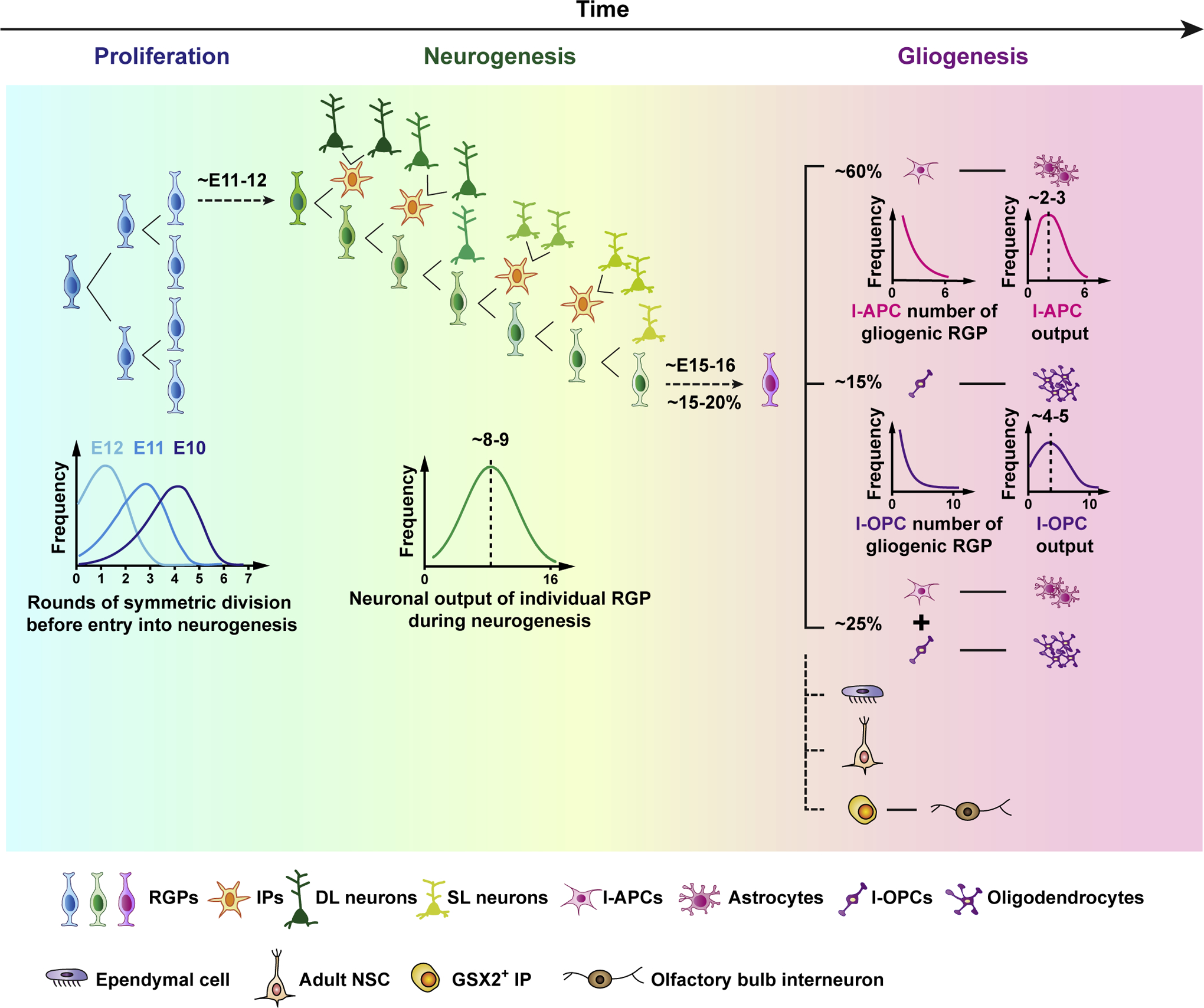
The cerebral cortex is a central structure in the mammalian brain that enables higher cognitive functions and intellectual skills. It is the hallmark of the mammalian nervous system with enormous complexity, consisting of a large number of neurons and glia that are diverse in morphology, molecular expression, biophysical properties, circuit connectivity and physiological function. Cortical neurons and glia are generated by neural progenitor cells during development. Ensuring the correct cell cycle kinetics, fate behavior and lineage progression of neural progenitor cells is essential to determine the number and types of neurons and glia in the cerebral cortex, which together constitute neural circuits for brain function. In this review, we discuss recent findings on mammalian cortical progenitor cell types and their lineage behaviors in generating neurons and glia, cortical evolution and expansion, and advances in brain organoid technology that allow the modeling of human cortical development under normal and disease conditions.


Conflict of interest statement

Conflict of interest statement

Nothing declared.

Figures

Figure 1.
Figure 1.. Highly organized behavior and lineage program of RGPs in the developing mouse cortex.
A schematic representation of mouse cortical RGP behavior and lineage progression. RGPs first amplify through a defined sequence of symmetric divisions before switching abruptly into a neurogenic phase (left). While the timing of the transition varies across the ensemble of RGPs, it is synchronized by division number within an individual RGP lineage in the amplification phase (left, lower). In the neurogenic phase, deep layer (DL) and then superficial layer (SL) excitatory neurons are generated through asymmetric RGP cell divisions, either directly or indirectly through IPs with more restricted division and neuronal output potentials (middle). Overall, individual RGPs give rise to a “unitary” number of neuronal output with a Gaussian-like distribution peaked at around 8–9 neurons (middle, lower). At the end of neurogenesis, a fraction (~20%) of RGPs switch abruptly into a gliogenic phase, giving rise to astrocytes and/or oligodendrocytes either directly or indirectly via fate-restricted intermediate astrocyte precursor cells (I-APCs) or intermediate oligodendrocyte precursor cells (I-OPCs) (right). While the total number of I-APCs and I-OPCs generated by individual gliogenic RGPs appears to be governed by defined probabilistic rules, the glial outputs of individual I-APCs and I-OPCs are relatively well-defined. A small number of RGPs also give rise to ependymal cells, adult ventricular-subventricular zone neural stem cells (i.e., type B1 cells), and GSX2+ IPs that give rise to GABAergic interneurons in the olfactory bulb.
Figure 2.
Figure 2.. Lineage variations of RGPs during neurogenesis and possible underlying mechanisms in the developing mouse cortex.
(a) Statistical variations in the neuronal output of individual RGPs labeled at ~E12.5 based on the Emx1-CreER;RCL-Gfp (left) and Emx1-CreER;MADM (right) reporter with respect to the number and laminar position reported by Llorca et al. and Gao et al. Laminar-restricted: DL, deep layer (5–6) neurons only; SL, superficial layer (2–4) neurons only; Translaminar: DL and SL. Note that, at this induction time, some RGPs will have yet to enter into neurogenesis, while others may have already completed a significant fraction of their neurogenic program (see Fig. 1). (b) Two possible sources of RGP neurogenesis lineage variations have been proposed: the heterogeneous neurogenesis configuration model (left; Llorca et al.), and the coherent neurogenesis program model (right; Gao et al.). In the former, based particularly on the existence of laminar-restricted clones, RGPs are proposed to be pre-specified into distinct subtypes, each generating a diverse but defined neuronal output. For example, RGPD generates DL neurons only, RGPS generates SL neurons only, and RGPSD generates both DL and SL neurons. During neurogenesis, different subtypes of RGPs with distinct division and neurogenesis behaviors are programmed to generate neurons independently across the RGP population, implying a model of high complexity and specificity. In particular, as detailed by Llorca et al., a model of some 27 parameters is required to capacity the repertoire of observed sublineage sizes and compositions pointing to a complex underlying program of transcriptional control. By contrast, in the latter model, RGPs are considered to be largely equipotent, unfolding the same neurogenic program, and bearing the same proliferative and neurogenic potential. Small variations in the total neuronal output from the ~8–9 neuron average reflect heterogeneity in the proliferative capacity of daughter IPs, while variations in the neuronal composition of individual RGP clones reflect the timing of entry into and exit neurogenesis, with DL neurons enriched in RGP lineages that enter into neurogenesis early, and SL neurons enriched in those that enter late. The coherent neurogenesis program model, which predicts well the progressive output of DL and SL neurons at both the clonal and population level with regard to the number and laminar position across the entire neurogenic phase, also has the benefit of parsimony, depending on few parameters linked to the measured distribution of entry times into, and the length of, the neurogenic phase.
Figure 3.
Figure 3.. Comparison of the architectonic structures of the embryonic and mature cortices in three representative species.
Schematic illustrations of the embryonic and adult cortical organizations in reptiles, rodents and primates. (a) In reptiles, neurogenesis occurs primarily by RGPs in the ventricular zone (VZ). Some TBR2+ IPs also exist in the VZ, but there is no obvious subventricular zone (SVZ). The mature cortex is a tri-laminar structure with one prominent excitatory neuron layer in the middle. (b) In rodents, in addition to the VZ consisting of RGPs, the SVZ is prominent with ample IPs. The extensive indirect neurogenesis through IPs contributes to the production of both DL and SL neurons, and the formation of a six-layer cortex. (c) In primates, in addition to the increase in RGPs in the VZ, the SVZ is further expanded, including IPs and oRGs that predominantly constitutes the inner (iSVZ) and outer subventricular zone (oSVZ), respectively. oRGs also provide additional scaffolds supporting and guiding neuronal migration. Variations in neural progenitor cell types and consequently neurogenesis, as well as cell migration, may lead to a distinct functional spatial organization of the cortex in different species. For example, the neural map of orientation selectivity in the visual cortex shows a ‘salt-and-pepper’ organization in the mouse, whereas it is ‘clustered’ in primates and other higher mammals. IZ, intermediate zone; CP, cortical plate.

Similar articles

Cited by

References

    1. Wonders CP, Anderson SA: The origin and specification of cortical interneurons. Nat Rev Neurosci 2006, 7:687–696. - PubMed
    1. Swinnen N, Smolders S, Avila A, Notelaers K, Paesen R, Ameloot M, Brone B, Legendre P, Rigo JM: Complex invasion pattern of the cerebral cortex bymicroglial cells during development of the mouse embryo. Glia 2013, 61:150–163. - PubMed
    1. Dzierzak E, Bigas A: Blood Development: Hematopoietic Stem Cell Dependence and Independence. Cell Stem Cell 2018, 22:639–651. - PubMed
    1. Florio M, Huttner WB: Neural progenitors, neurogenesis and the evolution of the neocortex. Development 2014, 141:2182–2194. - PubMed
    1. Haubensak W, Attardo A, Denk W, Huttner WB: Neurons arise in the basal neuroepithelium of the early mammalian telencephalon: a major site of neurogenesis. Proc Natl Acad Sci U S A 2004, 101:3196–3201. - PMC - PubMed

Publication types

LinkOut - more resources