Review
. 2020 Nov 19;9(11):2503.
doi: 10.3390/cells9112503.

Ribosomal Protein L10: From Function to Dysfunction

Affiliations
Review

Ribosomal Protein L10: From Function to Dysfunction

Daniela Pollutri et al. Cells. .

Abstract

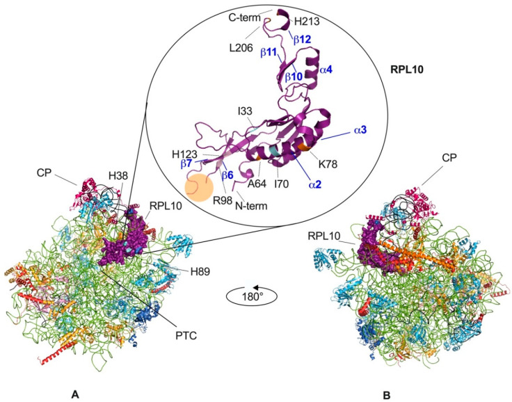
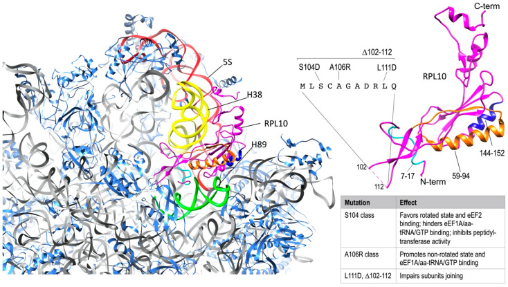
Eukaryotic cytoplasmic ribosomes are highly structured macromolecular complexes made up of four different ribosomal RNAs (rRNAs) and 80 ribosomal proteins (RPs), which play a central role in the decoding of genetic code for the synthesis of new proteins. Over the past 25 years, studies on yeast and human models have made it possible to identify RPL10 (ribosomal protein L10 gene), which is a constituent of the large subunit of the ribosome, as an important player in the final stages of ribosome biogenesis and in ribosome function. Here, we reviewed the literature to give an overview of the role of RPL10 in physiologic and pathologic processes, including inherited disease and cancer.

Keywords: RPL10; cancer; protein synthesis; rare disease; ribosome; ribosomopathy; translation.


Conflict of interest statement

The authors declare no conflict of interest.

Figures

Figure 2
Figure 2
Eukaryotic large ribosomal subunit and RPL10 mutations. Tetrahymena termophila 60S subunit, views of the 40S-exposed (A) and solvent-exposed (B) sides. RPL10 is in purple, 25S rRNA is in green, 5S is rRNA in black, and 5.8S is in carbon (visible in panel B). The localization of the central protuberance (CP), of the peptidyl-transferase center (PTC) and of other elements, which are significant for the present review are pinpointed in the 40S-exposed view. In the circle, the structure of the RPL10 protein is magnified, while clinically significant mutations are highlighted in different colors (pale pink for the mutations involved in T-ALL, light blue for the mutations involved in multiple myeloma, orange for the mutations involved in CNS-related abnormalities). The P-loop (between residues 102 and 112, orange circle) cannot be visualized because no ligand is present in this structure, as also explained in the main text. Some of the α helices and β sheets have been labeled, according to the numbering in Figure 1. The figure was generated using PyMOL (Schrodinger, LLC).
Figure 1
Figure 1
RPL10 and RPL10L (ribosomal protein L10-like gene) protein sequence alignment. The figure shows the multi-sequence alignment of RPL10 protein from Homo sapiens (P27635), Mus Musculus (Q6ZWV3), Saccharomyces cerevisiae (P41805), and Tetrahymena thermophila (Q235M8). In addition, the sequences of RPL10L from Homo sapiens (Q96L21) and Mus musculus (P86048) have been aligned. Sequences were aligned using Jalview program [17]. Each residue box in the alignment is assigned a color according to ClustalX color scheme. The higher the intensity of the color, the higher the conservation of the residue (white = not conserved residue) with respect to human RPL10. At the top of alignments the secondary structure of RPL10 from Tetrahymena thermophila is reported, which is visualized using PyMOL tool, as shown in Figure 2.
Figure 3
Figure 3
RPL10 ribosomal functions. On the left, Rpl10 in S. cervisiae 60S subunit (40S-exposed side); 25S rRNA is represented in grey (except for the colored portions, explained below) and RPLs other than Rpl10 are represented in cornflower blue. The regions of Rpl10 (magenta) that are relevant for interactions with rRNAs have been highlighted with different colors as follows: the portion including aminoacids 7-17 (light blue) is involved in contacting H89 in 25S rRNA (green); the portion including aminoacids 59-94 (orange) is placed between H38 in 25S rRNA (yellow) and H39 in 18S rRNA (not shown); the portion including aminoacids 144-152 (blue) is in close proximity of H89 (green); the C-terminal region is in contact with 5S rRNA (red). On the right side of the figure, Rpl10 structure is magnified and consistently colored. The unresolved sequence of the P-loop, with the different classes of mutations, as reported in the main text, has been added and the effects of the mutations in this site have been summarized in the side table. The figure was generated using UCSF Chimera [46].

Similar articles

Cited by

References

    1. Baßler J., Hurt E. Eukaryotic Ribosome Assembly. Annu. Rev. Biochem. 2019;88:281–306. doi: 10.1146/annurev-biochem-013118-110817. - DOI - PubMed
    1. Robledo S., Idol R.A., Crimmins D.L., Ladenson J.H., Mason P.J., Bessler M. The role of human ribosomal proteins in the maturation of rRNA and ribosome production. RNA. 2008;14:1918–1929. doi: 10.1261/rna.1132008. - DOI - PMC - PubMed
    1. Yamashita R., Suzuki Y., Takeuchi N., Wakaguri H., Ueda T., Sugano S., Nakai K. Comprehensive detection of human terminal oligo-pyrimidine (TOP) genes and analysis of their characteristics. Nucleic Acids Res. 2008;36:3707–3715. doi: 10.1093/nar/gkn248. - DOI - PMC - PubMed
    1. De la Cruz J., Karbstein K., Woolford J.L. Functions of Ribosomal Proteins in Assembly of Eukaryotic Ribosomes In Vivo. Annu Rev. Biochem. 2015;84:93–129. doi: 10.1146/annurev-biochem-060614-033917. - DOI - PMC - PubMed
    1. Peña C., Hurt E., Panse V.G. Eukaryotic ribosome assembly, transport and quality control. Nat. Struct. Mol. Biol. 2017;24:689–699. doi: 10.1038/nsmb.3454. - DOI - PubMed

Publication types