. 2020 Nov 19;21(22):8731.
doi: 10.3390/ijms21228731.

Suppression of Non-Random Fertilization by MHC Class I Antigens

Affiliations

Suppression of Non-Random Fertilization by MHC Class I Antigens

Junki Kamiya et al. Int J Mol Sci. .

Abstract

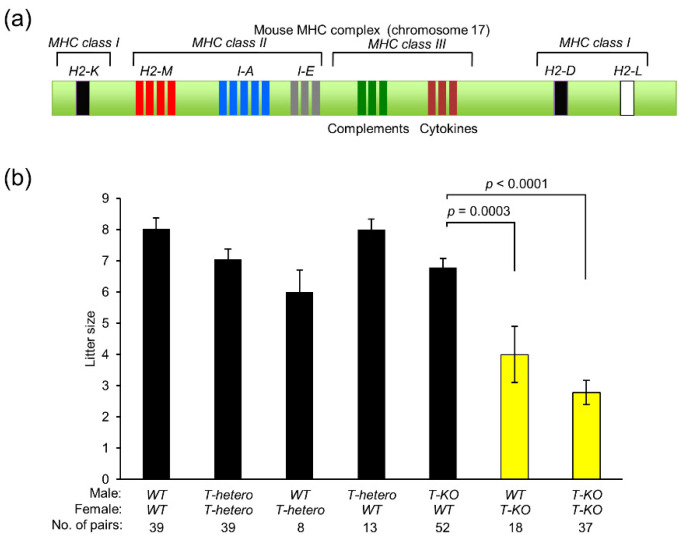
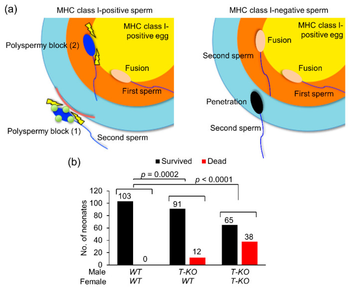
Hermaphroditic invertebrates and plants have a self-recognition system on the cell surface of sperm and eggs, which prevents their self-fusion and enhances non-self-fusion, thereby contributing to genetic variation. However, the system of sperm-egg recognition in mammals is under debate. To address this issue, we explored the role of major histocompatibility complex class I (MHC class I, also known as histocompatibility 2-Kb or H2-Kb and H2-Db in mice) antigens by analyzing H2-Kb-/-H2-Db-/-β2-microglobulin (β2M)-/- triple-knockout (T-KO) male mice with full fertility. T-KO sperm exhibited an increased sperm number in the perivitelline space of wild-type (WT) eggs in vitro. Moreover, T-KO sperm showed multiple fusion with zona pellucida (ZP)-free WT eggs, implying that the ability of polyspermy block for sperm from T-KO males was weakened in WT eggs. When T-KO male mice were intercrossed with WT female mice, the percentage of females in progeny increased. We speculate that WT eggs prefer fusion with T-KO sperm, more specifically X-chromosome-bearing sperm (X sperm), suggesting the presence of preferential (non-random) fertilization in mammals, including humans.

Keywords: MHC class I; non-random fertilization; polyspermy block; sex ratio.


Conflict of interest statement

The authors declare no conflict of interest.

Figures

Figure 1
Figure 1
Effect of major histocompatibility complex (MHC) class I antigens on mouse fertility. (a) Schematic representation of MHC genes on mouse chromosome 17. The MHC gene cluster contains MHC class I genes (H2-K, H2-D, and H2-L genes) and MHC class II genes (H2-M, and I-A and I-E subregions encoding glycoproteins). Each of H2-K, D, and L regions contain a single gene encoding an MHC class Iα chain. Each of I-A and I-E regions contains a single gene encoding an MHC class IIα chain, and one or more genes encoding MHC class IIβ chains. The MHC class III region contains genes encoding complement proteins, Heat shock proteins, tumor necrosis factor, and lymphotoxin. The H2-M region contains genes encoding class IIb proteins. (b) Litter size of progeny obtained from intercrossing between WT, T-hetero, and T-KO mice. Values are expressed as mean ± SEM.
Figure 2
Figure 2
Fertilization ability of T-KO sperm. (a) The fertilization success rate (%) after ovulated eggs were incubated with WT or T-KO sperm (103 sperm/mL) for 60 and 120 min. The extrusion of second polar body from the egg was designated as the success of fertilization. Parentheses indicate the number of eggs examined. (b) The fertilization success rate (%) after ovulated eggs were incubated with WT or T-KO sperm (104 sperm/mL) for 60, 90, and 120 min. The extrusion of second polar body was designated as the success of fertilization. Parentheses indicate the number of eggs examined. (c) The rate of polyspermy. Eggs penetrated by multiple sperm were defined as eggs that experienced polyspermy. Ovulated eggs were incubated with WT or T-KO sperm (103 sperm/mL or 104 sperm/mL) for 120 min. Parentheses indicate the number of eggs examined. (d) Experimental flow of sperm–egg fusion assay. (e) Representative images of WT eggs fused with T-KO and WT sperm. BF: bright-field microscopy; SN: sperm nucleus; EN: egg nucleus. Scale bar: 20 µm. (f) The number of sperm fused with an egg. Values are expressed as mean ± SEM. Parentheses indicate the number of eggs examined.
Figure 3
Figure 3
MHC class I antigens expressed on the sperm and progeny of MHC class I homozygous male and WT female. (a) Sex ratio in progeny of T-KO males and WT females (total neonates). (b) Sex ratio in progeny of T-KO males and WT females (litter size from 2 to 6). (c) Sex ratio in progeny dependent on the litter size. The values were analyzed by a chi-squared test. Values in parentheses indicate the number of neonates. (d) Immunoblotting with anti-H2-Kb polyAb in sperm extracts from T-hetero and T-KO males. CD9 and endogenously biotinylated proteins were detected as an internal control. (e) Immunocytochemical analysis of WT and T-KO sperm. The sperm were treated with anti-H2-Kb polyAb, and their nuclei were counterstained with DAPI. Scale bar: 10 µm. (f) Flow cytometric analysis of H2-Kb in the WT sperm. As negative controls, T-KO sperm were treated with anti-H2-Kb polyAb and WT sperm were treated with preimmune IgG. In histograms, the percentage of H2-Kb-positive sperm was determined by calculating the sperm number shifted to the right side, relative to the peak of WT sperm treated with preimmune IgG. (g) Hypothesis of male-biased fertilization. We assume that the sperm population from WT males could be divided into two groups: MHC-class-I-positive and MHC-class-I-negative population. MHC-class-I-negative population would be divided into two groups: X chromosome-bearing sperm (X sperm) and Y chromosome-bearing sperm (Y sperm). Under normal physiological conditions, MHC-class-I-committed random fertilization occurs, but MHC-class-I-negative sperm are not involved in fertilization. When the MHC-class-I-positive sperm population disappears, MHC-class-I-negative sperm, more specifically the X sperm, fertilizes MHC-class-I-positive eggs.
Figure 4
Figure 4
Schematic model of the role of MHC class I antigens in fertilization. (a) Fertilization by WT sperm. In the final process of fertilization, a sperm penetrates the zona pellucida and reaches the egg plasma membrane. After the sperm fuses with the WT egg plasma membrane, polyspermy block (1), a mechanism of polyspermy block is evoked, whereby extracellular release of egg materials is modified, which then blocks penetration by other sperm. Moreover, even if a second sperm arrives at the egg plasma membrane, polyspermy block (2), a second mechanism of polyspermy block occurs, due to which the sperm is unable to fuse with the egg plasma membrane. On the other hand, in fertilization with T-KO sperm, both mechanisms of polyspermy block are weak, and multiple sperm can penetrate and fuse with the WT egg plasma membrane. (b) Neonatal death. The number of dead neonates and the ones that survived was compared among three mating combinations: T-KO male and WT female, T-KO male and T-KO female, and WT male and WT female. The values were analyzed by a chi-squared test.

Similar articles

References

    1. Ben-Shlomo R. The molecular basis of allorecognition in ascidians. Bioessays. 2008;30:1048–1051. doi: 10.1002/bies.20848. - DOI - PubMed
    1. Harada Y., Sawada H. Allorecognition mechanisms during ascidian fertilization. Int. J. Dev. Biol. 2008;52:637–645. doi: 10.1387/ijdb.072544yh. - DOI - PubMed
    1. Fujii S., Kubo K., Takayama S. Non-self- and self-recognition models in plant self-incompatibility. Nat. Plants. 2016;2:16130. doi: 10.1038/nplants.2016.130. - DOI - PubMed
    1. Harada Y., Takagaki Y., Sunagawa M., Saito T., Yamada L., Taniguchi H., Shoguchi E., Sawada H. Mechanism of self-sterility in a hermaphroditic chordate. Science. 2008;320:548–550. doi: 10.1126/science.1152488. - DOI - PubMed
    1. Carlisle J.A., Swanson W.J. Molecular mechanisms and evolution of fertilization proteins. J. Exp. Zool. B Mol. Dev. Evol. 2020 doi: 10.1002/jez.b.23004. - DOI - PubMed

MeSH terms

Substances

LinkOut - more resources