. 2020 Nov 19;56(11):624.
doi: 10.3390/medicina56110624.

Diagnostic Performance of Contrast-Enhanced Ultrasound (CEUS) in the Evaluation of Solid Renal Masses

Affiliations

Diagnostic Performance of Contrast-Enhanced Ultrasound (CEUS) in the Evaluation of Solid Renal Masses

Thomas Geyer et al. Medicina (Kaunas). .

Abstract

Background: The present study aims to evaluate the diagnostic performance of contrast-enhanced ultrasound (CEUS) for discriminating between benign and malignant solid renal masses.

Methods: 18 patients with histopathologically confirmed benign solid renal masses (11 oncocytomas, seven angiomyolipomas) as well as 96 patients with confirmed renal cell carcinoma (RCC) who underwent CEUS followed by radical or partial nephrectomy were included in this single-center study. CEUS examinations were performed by an experienced radiologist (EFSUMB Level 3) and included the application of a second-generation contrast agent.

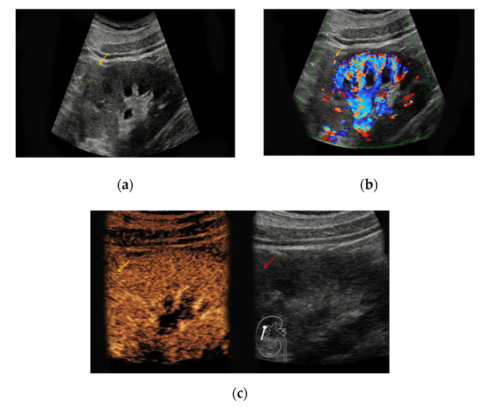
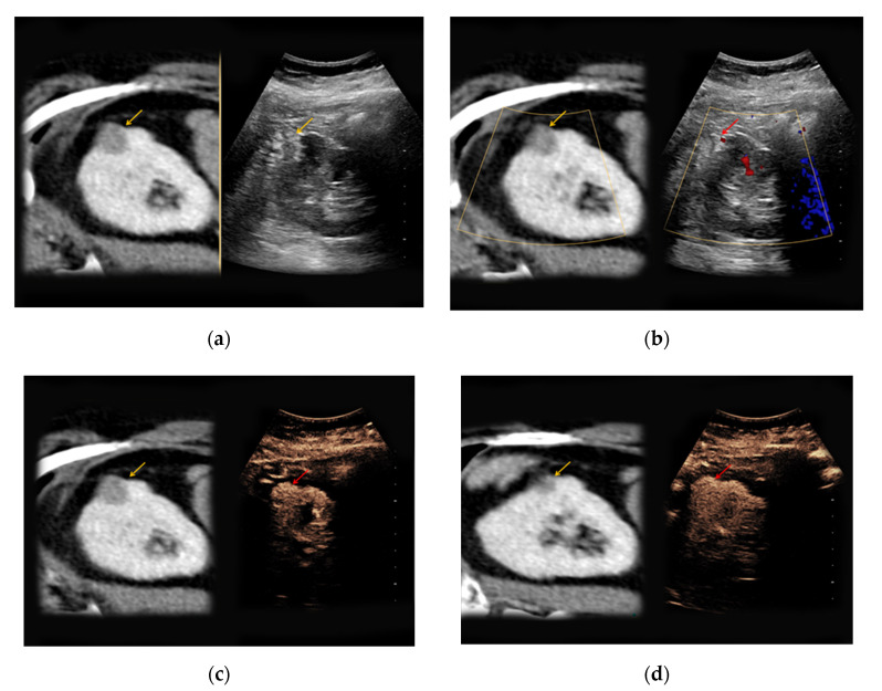
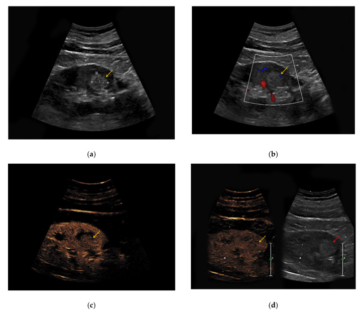
Results: Renal angiomyolipomas, oncocytomas, and renal cell carcinomas showed varying sonomorphological characteristics in CEUS. Angiomyolipomas showed heterogeneous echogenicity (57% hypo-, 43% hyperechoic), while all lesions showed rapid contrast-enhancement with two lesions also showing venous wash-out (29%). Notably, 9/11 oncocytomas could be detected in conventional ultrasound (64% hypo-, 9% hyper-, 9% isoechoic) and 2/11 only demarcated upon intravenous application of contrast agent (18%). All oncocytomas showed hyperenhancement in CEUS, venous wash-out was registered in 7/11 lesions (64%).

Conclusions: In line with the current state of knowledge, no specific sonomorphological characteristics allowing for accurate distinction between benign and malignant solid renal masses in CEUS could be detected in our study.

Keywords: CEUS; contrast-enhanced ultrasound; renal angiomyolipoma; renal cell carcinoma; renal oncocytoma; solid renal mass.


Conflict of interest statement

The authors declare no conflict of interest.

Figures

Figure 1
Figure 1
Renal oncocytoma in the right kidney with a diameter of 1.2 cm. (a) In native B-mode, the lesion presents as slightly hyperechoic (yellow arrow); (b) the lesion shows discrete vascularization in Doppler mode. (c) Upon intravenous application of SonoVue®, the lesion shows no signs of hyperenhancement.
Figure 2
Figure 2
An 81-year-old patient with an unclear lesion in the right kidney incidentally detected in contrast-enhanced computed tomography (CT). (a) In native B-mode, an inhomogeneously hyperechoic lesion with a diameter of 2.5 cm could be registered (right picture, yellow arrow). In the corresponding contrast-enhanced CT scan (a, left picture, yellow arrow), the lesion presents hypodense with only low contrast-enhancement; (b) no major vascularization could be visualized in Doppler mode (right picture, red arrow). (c) CEUS shows a rapid hyperenhancement of the lesion (right picture, red arrow); (d) no wash-out in the late phase was registered (right picture, red arrow). The patient underwent partial nephrectomy, the results of the histopathological analysis revealed benign renal angiomyolipoma.
Figure 3
Figure 3
(a) Hyperechoic renal lesion with a diameter of 1.6 cm visualized in native B-mode (yellow arrow); (b) only slight vascularization was registered in Doppler mode (yellow arrow). (c,d) The lesion did not show hyperenhancement during the early (c, yellow arrow) and late phase (d, yellow arrow). Histopathological results showed a clear-cell renal cell carcinoma.

Similar articles

Cited by

References

    1. Decastro G.J., McKiernan J.M. Epidemiology, clinical staging, and presentation of renal cell carcinoma. Urol. Clin. N. Am. 2008;35:581–592. doi: 10.1016/j.ucl.2008.07.005. - DOI - PubMed
    1. Chow W.H., Devesa S.S., Warren J.L. Rising incidence of renal cell cancer in the United States. JAMA. 1999;281:1628–1631. doi: 10.1001/jama.281.17.1628. - DOI - PubMed
    1. Jamis-Dow C.A., Choyke P.L., Jennings S.B. Small (< or = 3-cm) renal masses: Detection with CT versus US and pathologic correlation. Radiology. 1996;198:785–788. - PubMed
    1. Kang S.K., Chandarana H. Contemporary imaging of the renal mass. Urol. Clin. North Am. 2012;39:161–170. doi: 10.1016/j.ucl.2012.01.002. - DOI - PubMed
    1. Ljungberg B., Bensalah K., Canfield S. EAU guidelines on renal cell carcinoma: 2014 update. Eur. Urol. 2015;67:913–924. doi: 10.1016/j.eururo.2015.01.005. - DOI - PubMed