. 2020 Nov 19;9(11):3714.
doi: 10.3390/jcm9113714.

A One-Year Radiographic Healing Assessment after Endodontic Microsurgery Using Cone-Beam Computed Tomographic Scans

Affiliations

A One-Year Radiographic Healing Assessment after Endodontic Microsurgery Using Cone-Beam Computed Tomographic Scans

Sumi Kang et al. J Clin Med. .

Abstract

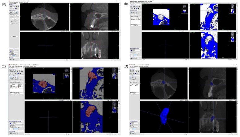
This study aimed to evaluate one-year radiographic healing after endodontic microsurgery using CBCT with modified PENN 3D criteria and to compare the outcome with results evaluated using Molven's criteria. A total of 107 teeth from 96 patients were evaluated one year after endodontic microsurgery by using CBCT scans with modified PENN 3D criteria and periapical radiographs with Molven's criteria. Both preoperative and postoperative lesion volumes were calculated using ITK-SNAP (free software). Radiographic healing assessment using periapical radiographs and CBCT images, and preoperative and postoperative lesion volume measurements were performed independently by two examiners. The assessment using Molven's criteria resulted in 75 complete healings, 18 incomplete healings, eight uncertain healings, and six unsatisfactory healings. Based on modified PENN 3D criteria, 64 teeth were categorized as complete healing, 29 teeth as limited healing, six teeth as uncertain healing, and eight teeth as unsatisfactory healing. With the one-year follow-up, CBCT scans showed a lower healing tendency than did periapical radiography. The volumes of apical radiolucency after the surgery were reduced by 77.7% on average at one-year follow up.

Keywords: apical surgery; cone-bean computed tomography (CBCT); one-year follow up; radiographic healing.


Conflict of interest statement

The authors declare no conflict of interest.

Figures

Figure 1
Figure 1
Lesion volume calculation using ITK-SNAP. (A) manual segmentation of a periapical defect in sagittal, axial and coronal views. (B) Selection of periapical defects using a grayscale value range selection tool. (C) Semiautomatic defect volume recognition and (D) reconstruction of 3D image on a lesion and calculation of the lesion volume.
Figure 2
Figure 2
(A) Examples of agreement cases. The images in each row were scored 1, 2, 3 and 4 resulted from both 2D and 3D assessments. (B) Examples of disagreement cases. The images at the first row were scored 1 from 2D assessment, but scored 2 from 3D assessment. The images at the second row were scored 3 from 2D assessment, but 4 from 3D assessment.

References

    1. Molven O., Halse A., Grung B. Observer strategy and the radiographic classification of healing after endodontic surgery. Int. J. Oral Maxillofac. Surg. 1987;16:432–439. doi: 10.1016/S0901-5027(87)80080-2. - DOI - PubMed
    1. Rud J., Andreasen J.O., Jensen J.E. Radiographic criteria for the assessment of healing after endodontic surgery. Int. J. Oral Surg. 1972;1:195–214. doi: 10.1016/S0300-9785(72)80013-9. - DOI - PubMed
    1. Song M., Nam T., Shin S.J., Kim E. Comparison of clinical outcomes of endodontic microsurgery: 1 year versus long-term follow-up. J. Endod. 2014;40:490–494. doi: 10.1016/j.joen.2013.10.034. - DOI - PubMed
    1. Von Arx T., Jensen S.S., Janner S.F.M., Hanni S., Bornstein M.M. A 10-year Follow-up Study of 119 Teeth treated with apical surgery and root-end filling with mineral trioxide aggregate. J. Endod. 2019;45:394–401. doi: 10.1016/j.joen.2018.12.015. - DOI - PubMed
    1. Lo Giudice R., Nicita F., Puleio F., Alibrandi A., Cervino G., Lizio A.S., Pantaleo G. Accuracy of periapical radiography and CBCT in endodontic evaluation. Int. J. Dent. 2018;2018:2514243. doi: 10.1155/2018/2514243. - DOI - PMC - PubMed

LinkOut - more resources