. 2020 Nov 19;8(4):107.
doi: 10.3390/toxics8040107.

An Embryonic Zebrafish Model to Screen Disruption of Gut-Vascular Barrier upon Exposure to Ambient Ultrafine Particles

Affiliations

An Embryonic Zebrafish Model to Screen Disruption of Gut-Vascular Barrier upon Exposure to Ambient Ultrafine Particles

Kyung In Baek et al. Toxics. .

Abstract

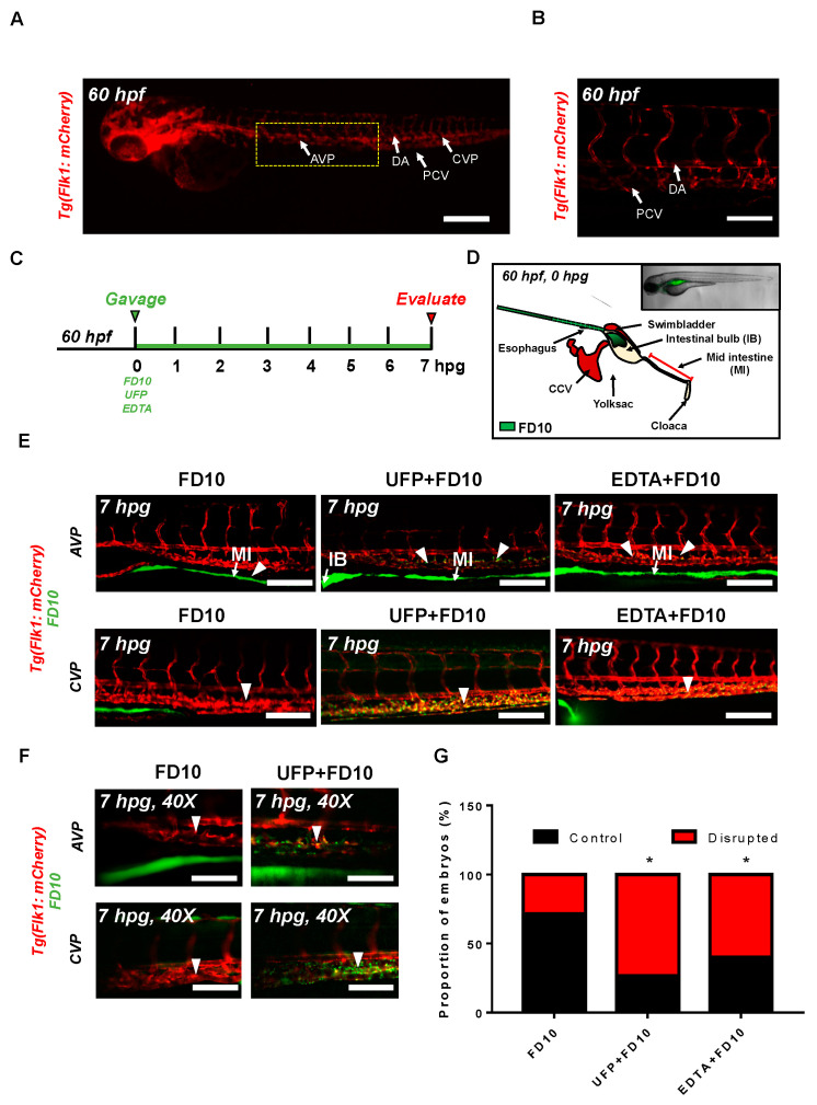
Epidemiological studies have linked exposure to ambient particulate matter (PM) with gastrointestinal (GI) diseases. Ambient ultrafine particles (UFP) are the redox-active sub-fraction of PM2.5, harboring elemental and polycyclic aromatic hydrocarbons from urban environmental sources including diesel and gasoline exhausts. The gut-vascular barrier (GVB) regulates paracellular trafficking and systemic dissemination of ingested microbes and toxins. Here, we posit that acute UFP ingestion disrupts the integrity of the intestinal barrier by modulating intestinal Notch activation. Using zebrafish embryos, we performed micro-gavage with the fluorescein isothiocynate (FITC)-conjugated dextran (FD10, 10 kDa) to assess the disruption of GVB integrity upon UFP exposure. Following micro-gavage, FD10 retained in the embryonic GI system, migrated through the cloaca. Conversely, co-gavaging UFP increased transmigration of FD10 across the intestinal barrier, and FD10 fluorescence occurred in the venous capillary plexus. Ingestion of UFP further impaired the mid-intestine morphology. We performed micro-angiogram of FD10 to corroborate acute UFP-mediated disruption of GVB. Transient genetic and pharmacologic manipulations of global Notch activity suggested Notch regulation of the GVB. Overall, our integration of a genetically tractable embryonic zebrafish and micro-gavage technique provided epigenetic insights underlying ambient UFP ingestion disrupts the GVB.

Keywords: Notch signaling; gut-vascular barrier; micro-gavage; ultrafine particles; zebrafish.


Conflict of interest statement

The authors declare no conflict of interest.

Figures

Figure 1
Figure 1
Acute ultrafine particulate matter (UFP) ingestion disrupts intestinal epithelial barrier integrity. Transgenic Tg(flk1: mCherry) zebrafish embryos at 60 hpf were micro-gavaged with FITC-conjugated dextran (FD10, 10 kDa). (A,B) Anatomy of endothelial vasculature in the Tg(flk1: mCherry) embryo. DA: dorsal aorta; PCV: posterior caudal vein; AVP: anterior venous capillary plexus; CVP: caudal vein capillary plexus; Scale bar: 32 μm. (C) Experimental design: At 2 dpf, embryos were randomly chosen for micro-gavage with FD10 solution with or without UFP or EDTA at 20 mM. Intestinal barrier integrity and translocation of FD10 to vascular endolumen (flk1+) were evaluated at 7 h post gavage (hpg). (D) A schematic representation of micro-gavage in an embryonic gastrointestinal (GI) tract. FD10 solution was micro-gavaged in the intestinal bulb without disrupting the esophagus, swim bladder and yolk sac. (E) Representative images of the AVP and CVP at 7 hpg. In FD10 gavaged-controls, FD10 remained only in the intestinal bulb and mid-intestine. In contrast, co-gavaging FD10 with UFP or ethylenediaminetetraacetic acid (EDTA) accumulated FD10 in the AVP and CVP (white arrow heads). Scale bar: 20 μm. (F) Magnified view of the AVP and CVP. Scale bar: 20 μm. (G) Percentage of embryos exhibiting endoluminal FD10 fluorescence (* p < 0.05 vs. FD10, n = 10 per group).
Figure 2
Figure 2
Acute UFP exposure disrupts maturation of embryonic GI tract. (A) Schematic representation of the embryonic GI tract at 7 hpg. The density of FD10 fluorescence in the mid-intestine was assessed to evaluate maturation of the GI tract (Red box). (B) Representative images of UFP-disrupted GI tract (white dashed box). Compared to FD10-gavaged controls, co-gavaging UFP, as denoted with endoluminal FD10 fluorescence (white arrow), altered morphology and systemically reduced the density of FD10 fluorescence in the mid-intestine (white arrowheads, n = 5 per group). Scale bar: 20 μm. (C) Schematic representations of sagittal and transverse views of the mid-intestine with and without UFP gavage. Acute UFP exposure in developing GI system retards maturation (red arrowheads). (D) Percentage of embryos exhibiting reduced FD10 density in the mid intestine (* p < 0.05 vs. FD10, n = 10 per group).
Figure 3
Figure 3
Micro-angiography via common cardinal vein (CCV) to mimic UFP gavage. (A) A schematic representation of micro-angiography via CCV to introduce FD10 to the microcirculatory system. (B) A representative image of the transgenic Tg(flk1: mCherry) embryo following FD10 injection to CCV. At 0 hpi, FD10 fluorescence was prominent at the injection site, CCV, heart, DA and PCV. Scale bar: 100 μm. (C,D) At 1 hpi, FD10 was distributed in the AVP and CVP, between the DA and PCV, mimicking UFP gavage-mediated effects (white arrowheads, n = 5 per group). Scale bar: 32 μm.
Figure 4
Figure 4
UFP exposure down-regulates mRNA expressions of endothelial tight junction (TJ) protein and the Notch target gene to disrupt the GVB. mRNA expressions of TJ proteins, including zonula occludens1 (Zo1), claudin 1 (Cldn1), and occludin 1 (Ocln1), and the Notch target genes, including Hairy and enhancer of split-1 (Hes1), were assessed in vitro by cultured human aortic endothelial cells (HAEC). (A) UFP exposure (25 μg·mL−1 for 6 h) inhibited Zo1 and Cldn1 mRNA expressions, whereas Ocln1 mRNA remained unchanged. While Iwr1 treatment (10 μM) diminished overall TJ mRNA expression, LiCl (20 mM) up-regulated Cldn1 and Ocln1 mRNA expression (* p < 0.05 vs. DMSO for Iwr1, H2O for LiCl, n = 3, ** p < 0.01, *** p < 0.001). (B) UFP exposure (25–50 μg·mL−1 for 6 h) down-regulated both TJ (Zo1 and Cldn1 mRNA) and Notch target genes (Hes1) mRNA expression in a dose-dependent manner (* p < 0.05 vs. H2O, n = 3). (C) Treatment of Adam10 inhibitor (5 μM) to inhibit Notch receptor activation down-regulated Cldn1, and Hes1 mRNA in a dose-dependent manner (* p < 0.05 vs. DMSO, n = 3, ** p < 0.01, *** p < 0.001) (D) Micro-gavage with Adam10 inhibitor promoted transmigration of FD10 to both AVP and CVP. Micro-injection of NICD mRNA restored UFP- and Adam10 inhibitor-mediated effect. (E) Percentage of embryos exhibiting endoluminal FD10 fluorescence (* p < 0.05 vs. FD10, n = 10 per group).
Figure 5
Figure 5
Schematic overviews of the proposed mechanisms.

References

    1. Nel A., Xia T., Madler L., Li N. Toxic potential of materials at the nanolevel. Science. 2006;311:622–627. doi: 10.1126/science.1114397. - DOI - PubMed
    1. Zhang Y., Schauer J.J., Shafer M.M., Hannigan M.P., Dutton S.J. Source apportionment of in vitro reactive oxygen species bioassay activity from atmospheric particulate matter. Environ. Sci. Technol. 2008;42:7502–7509. doi: 10.1021/es800126y. - DOI - PubMed
    1. Kaplan G. Air pollution and the inflammatory bowel diseases. Inflamm. Bowel Dis. 2011;17:1146–1148. doi: 10.1002/ibd.21449. - DOI - PubMed
    1. Beamish L.A., Osornio-Vargas A.R., Wine E. Air pollution: An environmental factor contributing to intestinal disease. J. Crohns Colitis. 2011;5:279–286. doi: 10.1016/j.crohns.2011.02.017. - DOI - PubMed
    1. Lomer M.C., Thompson R.P., Powell J.J. Fine and ultrafine particles of the diet: Influence on the mucosal immune response and association with Crohn’s disease. Proc. Nutr. Soc. 2002;61:123–130. doi: 10.1079/PNS2001134. - DOI - PubMed

LinkOut - more resources