. 2020 Nov 19;10(11):293.
doi: 10.3390/life10110293.

Anti-Inflammatory Effects of Fermented Lotus Root and Linoleic Acid in Lipopolysaccharide-Induced RAW 264.7 Cells

Affiliations

Anti-Inflammatory Effects of Fermented Lotus Root and Linoleic Acid in Lipopolysaccharide-Induced RAW 264.7 Cells

Sung Min Kim et al. Life (Basel). .

Abstract

Inflammation is a protective response of the innate immune system. However, aberrant inflammatory responses lead to various diseases. Lotus root, the edible rhizome of Nelumbo nucifera, is a popular traditional herbal medicine in East Asia. In a previous study, we reported that fermented lotus root (FLR) alleviated ethanol/HCl-induced gastric ulcers in rats by modulating inflammation-related genes. However, the mechanisms underlying the anti-inflammatory effects of FLR and its major constituent, linoleic acid (LA), are still largely unknown. In this study, we investigated the anti-inflammatory effects of FLR and LA on lipopolysaccharide (LPS)-induced inflammation in RAW 264.7 murine macrophages. We found that FLR inhibited LPS-induced expression of inflammatory mediators through down-regulation of NF-κB activity. Similarly, LA also attenuated LPS-induced inflammatory responses and reduced LPS-induced phosphorylation of proteins associated with NF-κB signaling, such as ERK, JNK, and p38. Overall, our results suggested that FLR and LA may effectively ameliorate inflammatory diseases.

Keywords: MAPK; NF-κB; anti-inflammatory effect; fermented lotus root.


Conflict of interest statement

The authors declare no conflict of interest.

Figures

Figure 1
Figure 1
Effects of fermented lotus root (FLR) on cell viability. Cell viability in (a) RAW 264.7 murine macrophages treated with 25 to 200 μg/mL FLR for 24 h. (b) RAW 264.7 cells co-treated with FLR and lipopolysaccharide (LPS, 1 μg/mL) for 24 h. Cell viability is expressed as a percentage of the vehicle control (VC). The data represent mean ± SEM, n = 3. *** p < 0.001 compared to LPS-only treated group.
Figure 2
Figure 2
Effects of FLR on nitric oxide (NO) production and immune gene expression. RAW 264.7 cells were co-treated with LPS (1 μg/mL) and FLR (25 to 200 μg/mL) for 24 h. (a) NO production in culture medium (bf) mRNA expression of (b) Nos2, (c) Ptgs2, (d) Tnf-α, (e) Il1b, and (f) Il6 quantified by RT-PCR. The data represent mean ± SEM, n = 3. * p < 0.05, ** p < 0.01, *** p < 0.001 compared to LPS-only treated group.3.3. Effects of FLR on the activation and nuclear translocation of NF-κB p65.
Figure 3
Figure 3
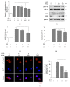
Effects of FLR on NF-κB signaling. (a) NF-κB promoter activity in transfected RAW 264.7 cells co-treated with LPS (1 µg/mL) and FLR (25 to 200 μg/mL) for 24 h. (bd) Western blot analysis of phosphorylated and unphosphorylated NF-κB and IκBα extracted from cells co-treated with 1 μg/mL LPS and 100, 200 μg/mL FLR for 1 h. The data represent mean ± SEM, n = 3. * p < 0.05, *** p < 0.001 compared to LPS-only treated group. (e) Representative images from immunofluorescence. The nucleus (blue color) was stained with 4′,6-Diamidino-2-phenylindole dihydrochloride (DAPI) and NF-κB p65 (red color) was stained with appropriate antibodies. Images were captured at 400× magnification. The scale bar indicates 20 μm. The scale bar indicates 20 μm. The number of cells with p65 nuclear translocation were counted for the statistical analysis in three random fields of each group.
Figure 4
Figure 4
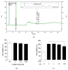
Chromatogram of linoleic acid (LA) and effects of LA on cell viability. (a) Chromatogram of LA was obtained using gas chromatography with flame ionization detector (GC-FID). (b) Cell viability in RAW 264.7 cells treated with different concentrations of LA (1, 10, and 100 μM) for 24 h. (c) Cell viability in RAW 264.7 cells co-treated with LPS (1 μg/mL) and LA for 24 h. Cell viability was measured by CCK-8 assay and expressed as a percentage compared to that of VC. The data represent mean ± SEM, n = 3, * p < 0.05 compared to LPS-only treated group.
Figure 5
Figure 5
Effects of LA on NO production and expression of immune genes. Cells were co-treated with LPS (1 μg/mL) and LA at the concentrations of 1, 10, and 100 μM for 24 h. (a) NO production in culture medium (bf) mRNA levels of immune genes Nos2(b), Ptgs2 (c), Tnf-α (d), Il1b (e), and Il6 (f). The data represent mean ± SEM, n = 3. * p < 0.05, ** p < 0.01, *** p < 0.001 compared to LPS-only treated group.
Figure 6
Figure 6
Effects of LA on NF-κB signaling. (a) NF-κB promoter activity in transfected RAW 264.7 cells co-treated with LPS and LA (1 to 100 μM) for 24 h. (bd) Western blot analysis of phosphorylated and unphosphorylated NF-κB and IκBα proteins extracted from cells co-treated with LPS (1 μg/mL) and LA (1 to 100 μM) for 1 h. The data represent mean ± SEM, n = 3. * p < 0.05, ** p < 0.01, *** p < 0.001 compared to LPS-only treated group. (e) Representative images from immunofluorescence. The nucleus (blue color) was stained with 4′,6-Diamidino-2-phenylindole dihydrochloride (DAPI) and NF-κB p65 (red color) was stained with appropriate antibodies. Images were captured at 400× magnification. The scale bar indicates 20 μm. The number of cells with p65 nuclear translocation were counted for the statistical analysis in three random fields of each group.
Figure 7
Figure 7
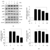
Effects of LA on phosphorylation of extracellular signal-regulated kinase (ERK), p38, and c-Jun N-terminal kinase (JNK). RAW 264.7 cells were co-treated with LPS (1 μg/mL) and 1 to 100 μM LA for 1 h. Western blot analysis of phosphorylated and unphosphorylated ERK, p38, and JNK proteins to evaluate MAPK activation The images above are representatives of triplicate experiments. The data represent mean ± SEM, n = 3. * p < 0.05, compared to LPS-only treated group.

Similar articles

Cited by

References

    1. De Lavor E.M., Fernandes A.W.C., de Andrade Teles R.B., Leal A., de Oliveira Junior R.G., Gama E.S.M., de Oliveira A.P., Silva J.C., de Moura Fontes Araujo M.T., Coutinho H.D.M., et al. Essential Oils and Their Major Compounds in the Treatment of Chronic Inflammation: A Review of Antioxidant Potential in Preclinical Studies and Molecular Mechanisms. Oxid. Med. Cell. Longev. 2018;2018:6468593. doi: 10.1155/2018/6468593. - DOI - PMC - PubMed
    1. Freigang S. The regulation of inflammation by oxidized phospholipids. Eur. J. Immunol. 2016;46:1818–1825. doi: 10.1002/eji.201545676. - DOI - PubMed
    1. Ara T., Nakatani S., Kobata K., Sogawa N., Sogawa C. The Biological Efficacy of Natural Products against Acute and Chronic Inflammatory Diseases in the Oral Region. Medicines. 2018;5 doi: 10.3390/medicines5040122. - DOI - PMC - PubMed
    1. Zerem E. Treatment of severe acute pancreatitis and its complications. World J. Gastroenterol. 2014;20:13879–13892. doi: 10.3748/wjg.v20.i38.13879. - DOI - PMC - PubMed
    1. Lu S., Wu D., Sun G., Geng F., Shen Y., Tan J., Sun X., Luo Y. Gastroprotective effects of Kangfuxin against water-immersion and restraint stress-induced gastric ulcer in rats: Roles of antioxidation, anti-inflammation, and pro-survival. Pharm. Biol. 2019;57:770–777. doi: 10.1080/13880209.2019.1682620. - DOI - PMC - PubMed