. 2020 Nov 19;9(11):963.
doi: 10.3390/pathogens9110963.

Rat Cytomegalovirus Virion-Associated Proteins R131 and R129 Are Necessary for Infection of Macrophages and Dendritic Cells

Affiliations

Rat Cytomegalovirus Virion-Associated Proteins R131 and R129 Are Necessary for Infection of Macrophages and Dendritic Cells

Iris K A Jones et al. Pathogens. .

Abstract

Cytomegalovirus (CMV) establishes persistent, latent infection in hosts, causing diseases in immunocompromised patients, transplant recipients, and neonates. CMV infection modifies the host chemokine axis by modulating chemokine and chemokine receptor expression and by encoding putative chemokine and chemokine receptor homologues. The viral proteins have roles in cellular signaling, migration, and transformation, as well as viral dissemination, tropism, latency and reactivation. Herein, we review the contribution of CMV-encoded chemokines and chemokine receptors to these processes, and further elucidate the viral tropism role of rat CMV (RCMV) R129 and R131. These homologues of the human CMV (HCMV)-encoded chemokines UL128 and UL130 are of particular interest because of their dual role as chemokines and members of the pentameric entry complex, which is required for entry into cell types that are essential for viral transmission and dissemination. The contributions of UL128 and UL130 to acceleration of solid organ transplant chronic rejection are poorly understood, and are in need of an effective in vivo model system to elucidate the phenomenon. We demonstrated similar molecular entry requirements for R129 and R131 in the rat cells, as observed for HCMV, and provided evidence that R129 and R131 are part of the viral entry complex required for entry into macrophages, dendritic cells, and bone marrow cells.

Keywords: chemokine receptors; chemokines; cytomegalovirus; dendritic cells; dissemination; viral entry.


Conflict of interest statement

The authors declare no conflict of interest. The funders had no role in the design of the study; in the collection, analyses, or interpretation of data; in the writing of the manuscript, or in the decision to publish the results.

Figures

Figure 1
Figure 1
Construction of RCMV R131 and R129 mutants and HiBiT tag fusions. A panel of RCMV recombinants containing mutations in R131 or R129 was created using BAC recombineering. Mutants and HiBiT containing viruses are color-coded to the data graphs. As depicted, R131 consists of one exon, whereas R129 contains two exons and an intron. Putative domains of R131 and R129 are labeled on the WT version of both genes. Both R131 and R129 contain predicted signal sequences (blue), CC-chemokine domains (grey), and acidic clusters predicted to be involved in pentamer formation, based off of homology with the essential regions of HCMV UL130 and UL128, respectively (green). Truncation mutants were created by deletion of residues, and the 2xSTOP mutants and R131 C36A mutation are shown in red.
Figure 2
Figure 2
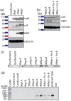
R131 is expressed with late viral gene expression kinetics. (a) Rat fibroblasts were infected with RCMV R131-HiBiT at a MOI = 1. Samples were washed with PBS and harvested in cell lysis buffer at 8, 24, and 48 hpi. Western blots for gB, RCMV IE, β-actin, and HiBiT (R131) were performed. (b) Rat fibroblasts were infected with RCMV R131-HiBiT at a MOI = 1 with or without foscarnet (0.5 mM) and samples were harvested in cell lysis buffer at 48 hpi. (c) Duplicate wells (A and B) of rat fibroblasts were infected with WT RCMV at an MOI = 1 with or without foscarnet (0.5 mM) and harvested in Trizol at 8, 24, and 48 hpi. RNA was isolated and Northern blots were performed probing for R131. (d) cDNA was made from RNA samples from (c) and reverse transcriptase PCR for R131 was performed. RCMV WT DNA was used as a positive control, water was used as the no template control (NTC). Size of select ladder bands are listed in base-pairs (bp).
Figure 3
Figure 3
R131 and R129 C’terminal regions are required for viral incorporation. (a) Viral incorporation of R129 and R131 was assessed for wild-type RCMV and viral mutants containing R129 and R131 HiBiT tags. The viruses were grown in rat fibroblasts. At the time of maximum cytopathic effect, supernatants were harvested and cellular debris was removed by centrifugation. Viral particles were then pelleted by ultracentrifugation over a 10% sorbitol gradient, and the resuspended virus pellet was additionally purified by banding over a discontinuous histodenz gradient. The banded virus was collected by ultracentrifugation, over a 10% sorbitol gradient. Purified viral particles were resuspended in PBS and equivalent quantities of viral particles were determined by blotting for gB. Lysates of rat fibroblasts infected with RCMV HiBiT-tagged mutants were harvested in cell lysis buffer with protease inhibitors. Equal quantities of protein, as determined by the BCA assay, were loaded onto the SDS-Page gels and detected by the HiBiT blot with LgBiT. HiBiT tagged R129 and R131 was detected in infected cell lysates (upper panel) for all viruses, including mutants. While WT HiBiT tagged R129 and R131 as well as the R131 C36A mutant were present in viral particles, the C’terminal deletion mutants were excluded from the purified viruses indicating that they were not incorporated (lower panel). (b) Viral particles were prepared as described in (a) for WT RCMV and the R129 and R131 HiBiT-tagged viruses. The samples were normalized to an amount of gB using Western blot (initial sample). Equal quantities of gB-containing viral particles were subjected to pull downs utilizing LgBiT-Halo Tag protein bound to Halo-Tag magnetic beads. The unbound fractions and bead bound fractions were analyzed by Western blotting for gB, R129, and HiBiT. (c–e) C’terminal deletions of R131 and R129 are not incorporated into viral particles. (c) Three different volumes of each virus preparation (7.5 μL, 3.75 μL, 1.875 μL) were assayed in triplicate against a standard curve by HiBiT lytic detection assay. Molecules of HiBiT per μL of virus preparation was determined using a commercially available standard HiBiT-tagged protein. (d) Viral DNA was extracted from 12.5 μL of each virus preparation and DNA was diluted 1:1000 and analyzed in triplicates by qPCR, using primers and probes, directed against the RCMV DNA polymerase. A standard curve of known concentration RCMV DNA was used to determine viral genome copies in each sample. (e) Molecules of HiBiT over viral genome copies in each sample was compared. Data from (c,d) were normalized per μL of the initial virus preparations.
Figure 4
Figure 4
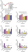
R131 and R129 are essential for entry into bone marrow, dendritic cells, and macrophages, but not fibroblasts or vascular smooth muscle cells. (a) Multistep growth curves were performed in triplicate wells by infecting rat fibroblasts with RCMV WT, R131 mutants, or R129 mutants at an MOI = 0.1. At 2 hpi, cells were washed three times with PBS and fresh medium was added to each well. Supernatant samples were collected at the time of infection and every 24 hpi until 120 hpi. The supernatants were titered by limiting dilution plaque assays in 24 well plates containing confluent monolayers of rat fibroblasts. The plates were fixed and stained after 7 days and viral titers were calculated. (bg) For entry assays, 96 well plates containing rat fibroblasts (b), vascular smooth muscle cells (c), SMG-derived epithelial cells (d), bone marrow cells (e), bone-marrow derived dendritic cells (f) and bone-marrow derived macrophages (g) were infected with RCMV WT, R131 mutants, or R129 mutants at a MOI = 0.5 in triplicate wells. At 20 hpi, the cells were fixed and stained with an α-RCMV IE polyclonal antibody and counterstained with DAPI, in order to count cell nuclei. Percent infection was determined by counting the number of IE positive cells divided by the number of cell nuclei. Percent of infection relative to WT virus was determined for each cell type. Data are representative of two independent experiments, each performed in triplicates. Statistical significance compared to infection levels with WT RCMV was determined for each viral mutant by one-way ANOVA, using Dunnett’s correction for multiple comparisons. ns = not significant, * p < 0.05, ** p < 0.01, *** p < 0.001, **** p < 0.0001.

Similar articles

Cited by

References

    1. Johansson I., Andersson R., Friman V., Selimovic N., Hanzen L., Nasic S., Nyström U., Sigurdardottir V. Cytomegalovirus infection and disease reduce 10-year cardiac allograft vasculopathy-free survival in heart transplant recipients. BMC Infect. Dis. 2015;15:582. doi: 10.1186/s12879-015-1321-1. - DOI - PMC - PubMed
    1. Ramanan P., Razonable R.R. Cytomegalovirus infections in solid organ transplantation: A review. Infect. Chemother. 2013;45:260–271. doi: 10.3947/ic.2013.45.3.260. - DOI - PMC - PubMed
    1. Orloff S.L., Hwee Y.-K.K., Kreklywich C., Andoh T.F., Hart E., Smith P.A., Messaoudi I., Streblow D.N. Cytomegalovirus latency promotes cardiac lymphoid neogenesis and accelerated allograft rejection in CMV Naive recipients. Am. J. Transplant. 2011;11:45–55. doi: 10.1111/j.1600-6143.2010.03365.x. - DOI - PMC - PubMed
    1. Melter M., McMahon G., Fang J., Ganz P., Briscoe D.M. Current understanding of chemokine involvement in allograft transplantation. Pediatr. Transplant. 1999;3:10–21. doi: 10.1034/j.1399-3046.1999.00023.x. - DOI - PubMed
    1. Russell M.E., Hancock W.W., Wallace A.F., Wyner L.R., Karnovsky M.J. Modulation of inflammatory activation pathways in the Lewis-to-F-344 rat chronic cardiac rejection model. Transplant. Proc. 1995;27:2100–2104. - PubMed

LinkOut - more resources