. 2020 Nov 19;8(11):1826.
doi: 10.3390/microorganisms8111826.

Proteomic Studies of the Biofilm Matrix including Outer Membrane Vesicles of Burkholderia multivorans C1576, a Strain of Clinical Importance for Cystic Fibrosis

Affiliations

Proteomic Studies of the Biofilm Matrix including Outer Membrane Vesicles of Burkholderia multivorans C1576, a Strain of Clinical Importance for Cystic Fibrosis

Lucrecia C Terán et al. Microorganisms. .

Abstract

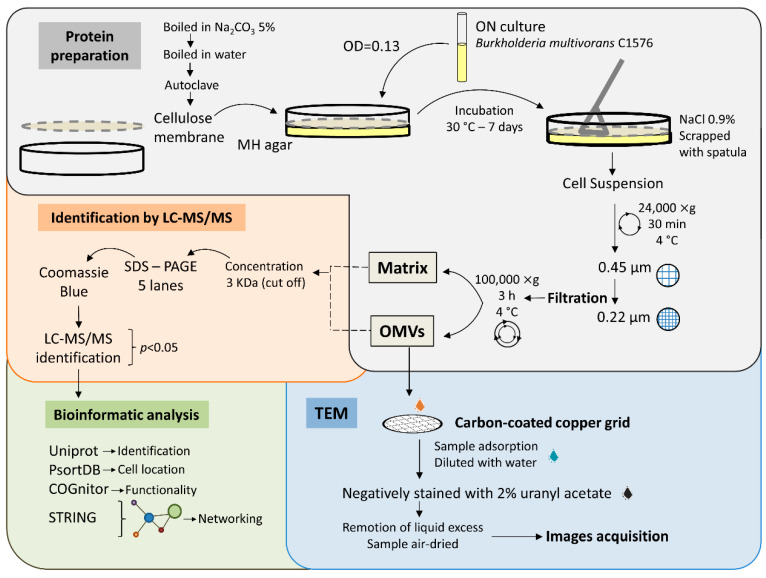
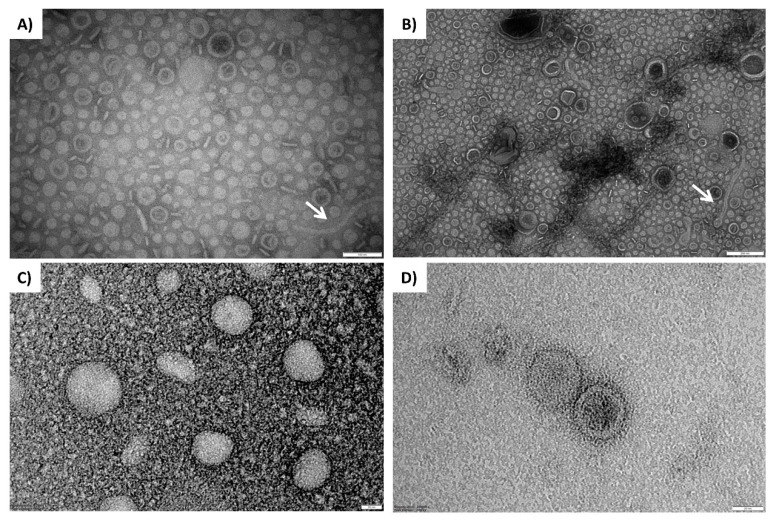
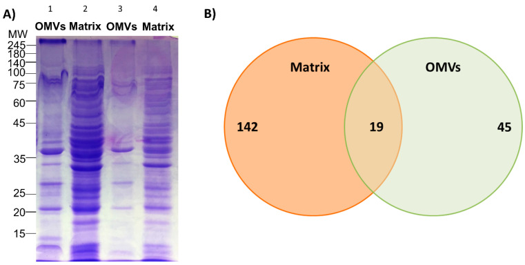
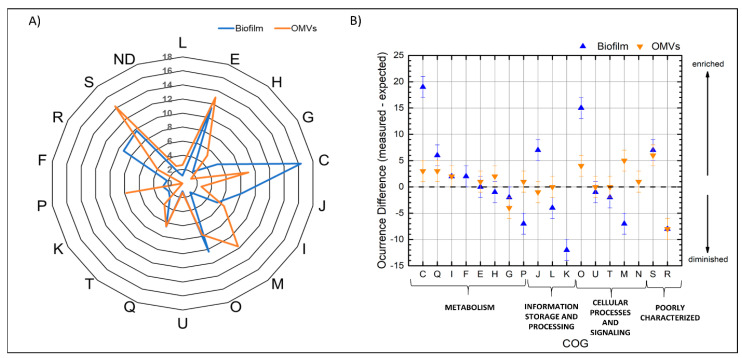
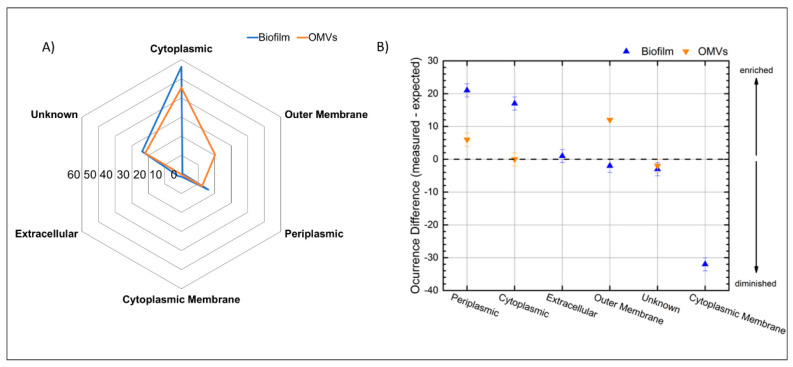
Biofilms are aggregates of microbial cells encased in a highly hydrated matrix made up of self-produced extracellular polymeric substances (EPS) which consist of polysaccharides, proteins, nucleic acids, and lipids. While biofilm matrix polysaccharides are unraveled, there is still poor knowledge about the identity and function of matrix-associated proteins. With this work, we performed a comprehensive proteomic approach to disclose the identity of proteins associated with the matrix of biofilm-growing Burkholderia multivorans C1576 reference strain, a cystic fibrosis clinical isolate. Transmission electron microscopy showed that B. multivorans C1576 also releases outer membrane vesicles (OMVs) in the biofilm matrix, as already demonstrated for other Gram-negative species. The proteomic analysis revealed that cytoplasmic and membrane-bound proteins are widely represented in the matrix, while OMVs are highly enriched in outer membrane proteins and siderophores. Our data suggest that cell lysis and OMVs production are the most important sources of proteins for the B. multivorans C1576 biofilm matrix. Of note, some of the identified proteins are lytic enzymes, siderophores, and proteins involved in reactive oxygen species (ROS) scavenging. These proteins might help B. multivorans C1576 in host tissue invasion and defense towards immune system assaults.

Keywords: Burkholderia multivorans; LC-LC/MS; biofilm; outer membrane vesicles; proteomic.


Conflict of interest statement

The authors declare no conflict of interest. The funders had no role in the design of the study; in the collection, analyses, or interpretation of data; in the writing of the manuscript; or in the decision to publish the results.

Figures

Figure 1
Figure 1
The general work scheme followed in the present work. Proteins from biofilm were separated into 2 fractions called Matrix and outer membrane vesicles (OMVs); both fractions were subjected to identification by LC-MS/MS and bioinformatics analysis. OMVs’ fraction was further analyzed by transmission electron microscope (TEM).
Figure 2
Figure 2
TEM visualization of OMVs produced by biofilm-growing B. multivorans C1576. The upper micrographs show TEM imaging of OMVs of the stock sample; elongated structures could represent contaminations with bacterial appendages (white arrows). The lower micrographs show TEM imaging of OMVs coming from the 1:50 dilution of the stock sample. Scale bars: 100 nm (A), 200 nm (B), 20 nm (C), and 20 nm (D).
Figure 3
Figure 3
(A) 12% SDS-PAGE protein profile of OMVs and the biofilm matrix produced by B. multivorans C1576; 15 µL of both samples were loaded at 1:2 (lane 1 and 2) and 1:5 (lane 3 and 4) dilution from stock concentration. (B) Venn diagram showing proteins of the biofilm matrix, the OMVs, and the ones that overlap between both samples.
Figure 4
Figure 4
(A) Percentage of the cluster of orthologous group (COG) functional categories of the identified proteins from the biofilm matrix and the OMVs. (B) Difference between the measured and the expected occurrence of the hypergeometric analysis. The positives values represent the enrichment of certain categories while the decrease of certain functional categories is represented by negatives values; when the expected value is identical to the measured one it is represented as zero. Letters represent the functional categories as follows: L: DNA replication, recombination and repair; E: metabolism and transport of amino acids; H: metabolism and transport of coenzymes; G: metabolism and transport of carbohydrates; C: production and conversion of energy; J: transcription; I: lipidic metabolism; M: Cell wall structure, biogenesis and outer membrane; O: posttranslational modifications; U: Intracellular trafficking, secretion, and vesicular transport; T: mechanisms of signal transduction; Q: metabolism and transport of nucleotides; P: transport of inorganic ions; N: cell motility; K: translation including ribosomes biogenesis; F: metabolism and transport of nucleotides; R: general prediction function only; S: function unknown; ND: not determined.
Figure 5
Figure 5
(A) Percentage of the subcellular localization of the identified proteins from the biofilm matrix and the OMVs. (B) Difference between measured and expected occurrence for the proteins of each subcellular localization. Zero represents no differences in occurrence and separates potentially enriched categories (positive values) from the potentially reduced categories (negative values).
Figure 6
Figure 6
Protein–protein interaction network of the identified proteins of B. multivorans C1576 in the biofilm matrix. The circles highlight the proteins related with functional categories. C: energy production and conversion, J: translation, O: post-translational modification, E: transport and metabolism of aminoacids, P: transport and metabolism of inorganic ions, I: metabolism of lipids, and H: transport and metabolism of coenzymes. The proteins are represented by nodes, while the interactions among them are represented by edges. The strength of interactions is proportional to line thickness. The network was constructed with STRING v11.
Figure 7
Figure 7
Protein–protein interaction network of the identified proteins of B. multivorans C1576 in the OMVs. The circles highlight the proteins related to functional categories. O: post-translational modification, E: transport and metabolism of aminoacids, I: metabolism of lipids, H: transport and metabolism of coenzymes, and M: cell wall structure, biogenesis and outer membrane. The proteins are represented by nodes while the interactions between them by edges. The strength of interactions is proportional to line thickness. The network was constructed with STRING v11.

References

    1. Costerton J.W., Lewandowski Z., Caldwell D.E., Korber D.R., Lappin-Scott H.M. Microbial biofilms. Annu. Rev. Microbiol. 1995;49:711–745. doi: 10.1146/annurev.mi.49.100195.003431. - DOI - PubMed
    1. O’Toole G., Kaplan H.B., Kolter R. Biofilm formation as microbial development. Annu. Rev. Microbiol. 2000;54:49–79. doi: 10.1146/annurev.micro.54.1.49. - DOI - PubMed
    1. Allison D.G. The biofilm matrix. Biofouling. 2003;19:139–150. doi: 10.1080/0892701031000072190. - DOI - PubMed
    1. Branda S.S., Vik S., Friedman L., Kolter R. Biofilms: The matrix revisited. Trends Microbiol. 2005;13:20–26. doi: 10.1016/j.tim.2004.11.006. - DOI - PubMed
    1. Fong J.N.C., Yildiz F.H. Biofilm matrix proteins. Microbiol. Spectr. 2015;3:1–27. doi: 10.1128/microbiolspec.MB-0004-2014. - DOI - PMC - PubMed

LinkOut - more resources