. 2020 Nov 23;21(1):234.
doi: 10.1186/s12881-020-01176-x.

Mice lacking global Stap1 expression do not manifest hypercholesterolemia

Affiliations

Mice lacking global Stap1 expression do not manifest hypercholesterolemia

Babunageswararao Kanuri et al. BMC Med Genet. .

Abstract

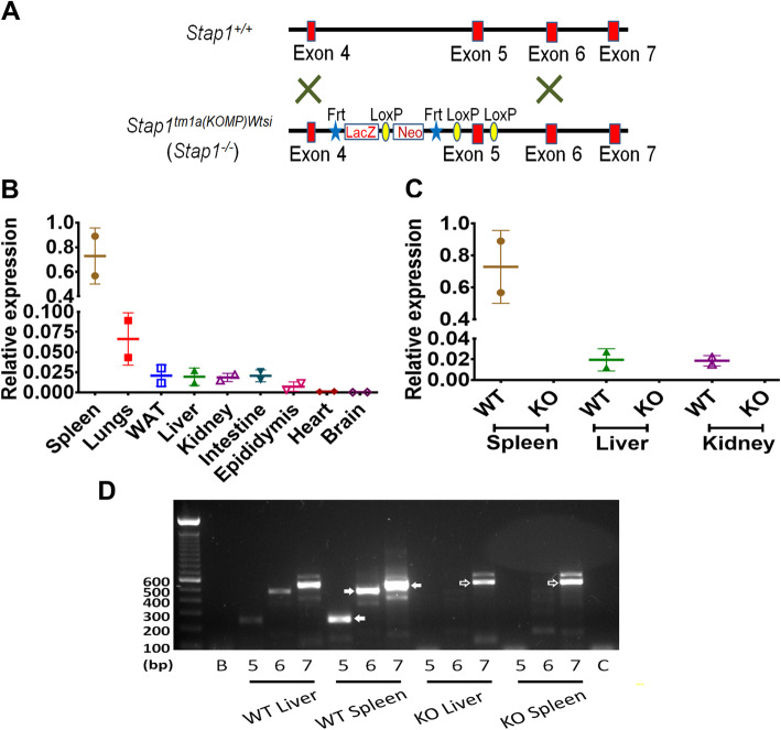
Background: Autosomal dominant familial hypercholesterolemia (ADH; MIM#143890) is one of the most common monogenic disorders characterized by elevated circulatory LDL cholesterol. Initial studies in humans with ADH identified a potential relationship with variants of the gene encoding signal transducing adaptor family member protein 1 (STAP1; MIM#604298). However, subsequent studies have been contradictory. In this study, mice lacking global Stap1 expression (Stap1-/-) were characterized under standard chow and a 42% kcal western diet (WD).

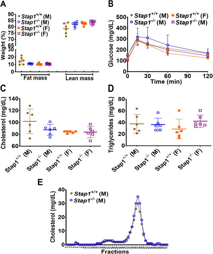
Methods: Mice were studied for changes in different metabolic parameters before and after a 16-week WD regime. Growth curves, body fats, circulatory lipids, parameters of glucose homeostasis, and liver architecture were studied for comparisons.

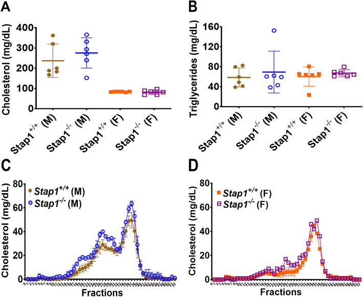
Results: Surprisingly, Stap1-/- mice fed the 16-week WD demonstrated no marked differences in any of the metabolic parameters compared to Stap1+/+ mice. Furthermore, hepatic architecture and cholesterol content in FPLC-isolated lipoprotein fractions also remained comparable to wild-type mice.

Conclusion: These results strongly suggest that STAP1 does not alter lipid levels, that a western diet did not exacerbate a lipid disorder in Stap1 deficient mice and support the contention that it is not causative for hyperlipidemia in ADH patients. These results support other published studies also questioning the role of this locus in human hypercholesterolemia.

Keywords: Autosomal dominant familial hypercholesterolemia; B-cells; Familial hypercholesterolemia 4; Fast performance liquid chromatography; STAP1; Western diet.


Conflict of interest statement

The authors declare no competing interests.

Figures

Fig. 1
Fig. 1
Verification of global deletion of Stap1 gene expression. Panel a shows the gene targeting details for the generation of Stap1−/− mice. This construct is a ‘knockout-first’, which can be converted to a conditional, upon deletion of the LacZ-Neo cassette. The mice used in this study remain as knockout and retain this cassette. A tissue survey of Stap1 mRNA was performed using qPCR in tissues collected from Stap1+/+ mice (panel b, n = 2). Note that compared to a house-keeping gene expression (see Methods), only spleen and lung showed some expression with very low levels detected in other tissues. Stap1−/− mice (KO) showed no detectable qRT-PCR products (panel c, n = 2–3) compared to wild-type mice (WT) in spleen, liver or kidney. To further confirm a lack of legitimate transcript expression, RT-PCR analysis of exon splicing for exons 2–5, 2–6, and 2–7 in liver and spleen was performed (panel d). The labels ‘5, 6, and 7’ for each tissue indicates the location of the reverse primers targeting exons 5, 6, or 7 respectively, used with a forward primer located in exon 2. Spleen mRNA from WT tissues amplified the correct expected size of product and size increments (exon 2–5; 251 bp, exon 2–6; 482 bp, and exon 2–7; 536 bp indicated by filled arrowheads). No products were noted when exons 2–5, or 2–6 were used for KO spleen mRNA. A slightly larger product was noted for exons 2–7 (indicated by open arrowhead), though on sequencing this was found to be a non-specific product from mis-priming (see Text). Error bars denote ±1 SD
Fig. 2
Fig. 2
Baseline metabolic parameters in chow-fed Stap1−/− and control mice. Panel a shows the starting body composition of mice at age 5–6 weeks on a chow diet (n = 6–7). Starting lean or fat masses between wildtype and knockout mice were comparable. Glucose tolerance (Panel b, n = 6), plasma cholesterol (Panel c, n = 6), triglycerides (Panel d, n = 6) were also comparable and showed no statistical differences. Panel e shows the FPLC cholesterol profiles in 10–11 weeks old male Stap1+/+ and Stap1−/− mice and was also indistinguishable. Note that the knockout mice do not show any differences in the VLDL (fractions 3–6), or LDL (fractions 20–28) ranges. Error bars denote ±1 SD
Fig. 3
Fig. 3
Effect of western diet on growth parameters and body composition in mice lacking Stap1 gene. Panel a shows the growth curves in wild-type and knockout mice and no statistical differences were discernable in the patterns of weight gain on the high caloric diet (n = 6). Neither the percent fat or lean mass were altered in Stap1−/− mice compared to age and sex-matched wildtype mice on a western diet (panel b, n = 6). No changes in weights of liver, spleen or kidneys were seen in male (panel c, n = 4–6) or female mice (panel d, n = 5–6) were noted. No changes in glucose tolerance tests were noted between knockout and wildtype controls either (panel e, n = 6). In case of male Stap1−/− mice, kidney weights of only 2 mice were considered for analysis
Fig. 4
Fig. 4
Effect of 16 weeks of western diet (WD) on plasma lipid profiles. Panel a shows the plasma cholesterol and panel b the triglyceride levels in wildtype and knockout mice (n = 6 per group). Female mice (whether wild-type or knockout) seem to be resistant to a WD-induced hypercholesterolemia (panel a), but this may be a feature of the mouse strain background. FPLC cholesterol profiles of the plasma (panel c, males, panel d, females) showed that the diet in males led to a dyslipidemic profile, but loss of Stap1 did not affect these patterns. Error bars denote ±1 SD
Fig. 5
Fig. 5
Effect of western diet (WD) on liver morphology and lipids. Assessment of liver histology by hematoxylin and eosin staining showed a fatty liver in all mice, irrespective of Stap1 status or sex (panel a and b). This was confirmed by analyses of liver cholesterol (panel c, n = 6 per group in males and 5 per group in females) and triglyceride (panel d, n = 6 per group in males and 5 per group in females) but loss of Stap1 does not affect these parameters. Error bars denote ±1 SD

Similar articles

Cited by

References

    1. Ezhov M, Bazhan S, Ershova A. Clinical guidelines for familial hypercholesterolemia. J Atherosclerosis Dyslipidemias. 2019;1:5–43.
    1. Goldberg AC, Hopkins PN, Toth PP, Ballantyne CM, Rader DJ, Robinson JG, Daniels SR, Gidding SS, de Ferranti SD, Ito MK, et al. Familial hypercholesterolemia: screening, diagnosis and management of pediatric and adult patients: clinical guidance from the National Lipid Association Expert Panel on familial hypercholesterolemia. J Clin Lipidol. 2011;5(3 Suppl):S1–S8. doi: 10.1016/j.jacl.2011.04.003. - DOI - PubMed
    1. Kanehisa M, Sato Y, Furumichi M, Morishima K, Tanabe M. New approach for understanding genome variations in KEGG. Nucleic Acids Res. 2019;47(D1):D590–D595. doi: 10.1093/nar/gky962. - DOI - PMC - PubMed
    1. Iacocca MA, Hegele RA. Recent advances in genetic testing for familial hypercholesterolemia. Expert Rev Mol Diagn. 2017;17(7):641–651. doi: 10.1080/14737159.2017.1332997. - DOI - PubMed
    1. Gidding SS, Champagne MA, de Ferranti SD, Defesche J, Ito MK, Knowles JW, McCrindle B, Raal F, Rader D, Santos RD, et al. The agenda for familial hypercholesterolemia: a scientific statement from the American Heart Association. Circulation. 2015;132(22):2167–2192. doi: 10.1161/CIR.0000000000000297. - DOI - PubMed

Publication types