. 2020 Nov 23;13(1):159.
doi: 10.1186/s13041-020-00698-y.

Focal loss of the paranodal domain protein Neurofascin155 in the internal capsule impairs cortically induced muscle activity in vivo

Affiliations

Focal loss of the paranodal domain protein Neurofascin155 in the internal capsule impairs cortically induced muscle activity in vivo

Kazuo Kunisawa et al. Mol Brain. .

Abstract

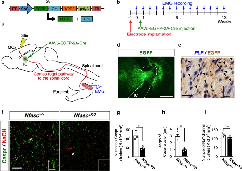
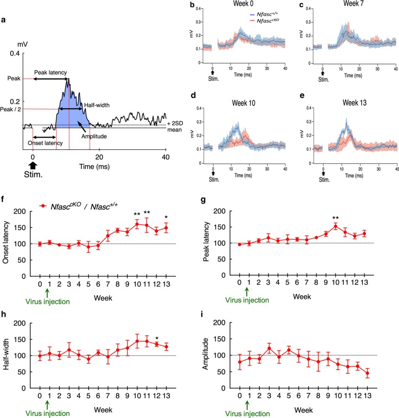
Paranodal axoglial junctions are essential for rapid nerve conduction and the organization of axonal domains in myelinated axons. Neurofascin155 (Nfasc155) is a glial cell adhesion molecule that is also required for the assembly of these domains. Previous studies have demonstrated that general ablation of Nfasc155 disorganizes these domains, reduces conduction velocity, and disrupts motor behaviors. Multiple sclerosis (MS), a typical disorder of demyelination in the central nervous system, is reported to have autoantibody to Nfasc. However, the impact of focal loss of Nfasc155, which may occur in MS patients, remains unclear. Here, we examined whether restricted focal loss of Nfasc155 affects the electrophysiological properties of the motor system in vivo. Adeno-associated virus type5 (AAV5) harboring EGFP-2A-Cre was injected into the glial-enriched internal capsule of floxed-Neurofascin (NfascFlox/Flox) mice to focally disrupt paranodal junctions in the cortico-fugal fibers from the motor cortex to the spinal cord. Electromyograms (EMGs) of the triceps brachii muscles in response to electrical stimulation of the motor cortex were successively examined in these awake mice. EMG analysis showed significant delay in the onset and peak latencies after AAV injection compared to control (Nfasc+/+) mice. Moreover, EMG half-widths were increased, and EMG amplitudes were gradually decreased by 13 weeks. Similar EMG changes have been reported in MS patients. These findings provide physiological evidence that motor outputs are obstructed by focal ablation of paranodal junctions in myelinated axons. Our findings may open a new path toward development of a novel biomarker for an early phase of human MS, as Nfasc155 detects microstructural changes in the paranodal junction.

Keywords: Electromyogram; Motor system; Multiple sclerosis; Neurofascin155; Paranodal junction.


Conflict of interest statement

The authors declare that they have no competing interests.

Figures

Fig. 1
Fig. 1
AAV-Cre-induced loss of paranodal junction in the internal capsule. a Schematic diagrams showing the design of the AAV-EGFP-2A-Cre constructs. The 2A peptide is cleaved just after translation, and EGFP and Cre recombinase are expressed independently. b, c Time course and schematic drawing of our experiments. To examine whether site-directed loss of paranodal junctions causes a delay in cortically evoked electromyograms (EMGs), a pair of bipolar stimulating electrodes was chronically implanted into the motor cortex (MCx), and EMG recording electrodes were placed in the triceps brachii muscles contralateral to the cortical stimulation electrodes in 6-week-old NfasccKO and Nfasc+/+ mice. AAV5-EGFP-2A-Cre was injected into the internal capsule (IC) of the mice at week 0. Evoked EMGs by cortical stimulation (Stim.) were recorded every week before (week 0) and after (from week 1 to week 13) AAV injection. d Representative micrograph of AAV-mediated EGFP expression (green) in the internal capsule 12 weeks after AAV injection. White dotted lines indicate the boundaries of the internal capsule (IC). Scale bar 250 μm. e Doubly staining by in situ hybridization for PLP mRNA (blue) and immunostaining with an anti-GFP antibody (brown) in the IC 12 weeks after AAV injection. Arrowheads indicate double positive cells for both PLP mRNA and GFP. The inset is a magnified view of the cell indicated by the arrow. Scale bar 50 μm (25 μm in inset). f Immunofluorescence staining with an anti-Caspr (green) and anti-Na+ channel (red) antibodies in sections containing the IC of Nfasc+/+ (left) or NfasccKO (right) mice 12 weeks after AAV injection. Each inset shows a magnified view of the immunolabeling indicated by the arrow. Scale bar 20 μm (5 μm in inset). Quantification of the number (g) and length (h) of Caspr-positive paranodes in Nfasc+/+ and NfasccKO mice injected with AAV5-EGFP-2A-Cre vector (Student’s t test, number [t = 4.623, df = 4, **p < 0.01], length [t = 5.319, df = 4, **p < 0.01]; n = 3 mice each). i Quantification of the number of Na+ channel-positive nodes in Nfasc+/+ and NfasccKO mice injected with AAV5-EGFP-2A-Cre vector (Student’s t test, t = 1.504, df = 4, p = 0.2069; n = 3 mice each). All data are shown as mean ± SEM
Fig. 2
Fig. 2
Cortically evoked EMGs in NfasccKO and Nfasc+/+ mice injected with AAV5-EGFP-2A-Cre. a Representative EMGs in response to electrical stimulation (0.6 mA, 100 μs duration, single pulse, 1400 ms interval, averaged 100 times) of the motor cortex in Nfasc+/+ mice 12 weeks after AAV injection. Onset latency, peak latency and half-width of the EMGs are indicated. Amplitude of the EMGs is defined as the area over the mean during the response exceeding the level of the mean + 2SD (blue area, see also “Materials and methods”). Cortically evoked population EMGs of Nfasc+/+ (n = 5, blue line) and NfasccKO (n = 7, red line) at week 0 (b), 7 (c), 10 (d) and 13 (e). The light-shaded colors represent ± SEM. Ratio (%) of onset latency (f), peak latency (g), half-width (h) and amplitude (i) of cortically evoked EMGs in NfasccKO mice compared to those of Nfasc+/+mice before (week 0) and after (from week 1 to week 13) AAV injection (two-way ANOVA followed by Sidak multiple comparison test; onset latency (genotype [F (1,137) = 21.41, **p < 0.01], week [F (13,137) = 2.480, **p < 0.01], genotype × week [F (13,137) = 2.480, **p < 0.01]), peak latency (genotype [F (1,125 = 20.19, **p < 0.01), week [F (13,125) = 1.179, p = 0.3024], genotype × week [F (13,125) = 1.179, p = 0.3024]], half-width (genotype [F (1,127) = 9.162, **p < 0.01], week [F (13,127) = 0.8992, p = 0.5558], genotype × week [F (13,127) = 0.9034, p = 0.5515]), amplitude (genotype [F (1,126) = 3.182, p = 0.0769], week [F (13,126) = 0.4220, p = 0.9595], genotype × week [F (13,126) = 0.4221, p = 0.9594])). All data are expressed as mean ± SEM

Similar articles

Cited by

References

    1. Aschauer DF, Kreuz S, Rumpel S. Analysis of transduction efficiency, tropism and axonal transport of AAV serotypes 1, 2, 5, 6, 8 and 9 in the mouse brain. PLoS ONE. 2013;8:e76310. doi: 10.1371/journal.pone.0076310. - DOI - PMC - PubMed
    1. Baumann N, Pham-Dinh D. Biology of oligodendrocyte and myelin in the mammalian central nervous system. Physiol Rev. 2001;81:871–927. doi: 10.1152/physrev.2001.81.2.871. - DOI - PubMed
    1. Bhat MA, Rios JC, Lu Y, Garcia-Fresco GP, Ching W, St Martin M, Li J, Einheber S, Chesler M, Rosenbluth J, Salzer JL, Bellen HJ. Axon-glia interactions and the domain organization of myelinated axons requires neurexin IV/Caspr/Paranodin. Neuron. 2001;30:369–383. doi: 10.1016/S0896-6273(01)00294-X. - DOI - PubMed
    1. Buttermore ED, Thaxton CL, Bhat MA. Organization and maintenance of molecular domains in myelinated axons. J Neurosci Res. 2013;91:603–622. doi: 10.1002/jnr.23197. - DOI - PMC - PubMed
    1. Calabrese M, Agosta F, Rinaldi F, Mattisi I, Grossi P, Favaretto A, Atzori M, Bernardi V, Barachino L, Rinaldi L, Perini P, Gallo P, Filippi M. Cortical lesions and atrophy associated with cognitive impairment in relapsing-remitting multiple sclerosis. Arch Neurol. 2009;66:1144–1150. doi: 10.1001/archneurol.2009.174. - DOI - PubMed

Publication types

LinkOut - more resources