Review
. 2020 Nov 23;13(1):588.
doi: 10.1186/s13071-020-04445-z.

The life-cycle of Toxoplasma gondii reviewed using animations

Affiliations
Review

The life-cycle of Toxoplasma gondii reviewed using animations

Márcia Attias et al. Parasit Vectors. .

Abstract

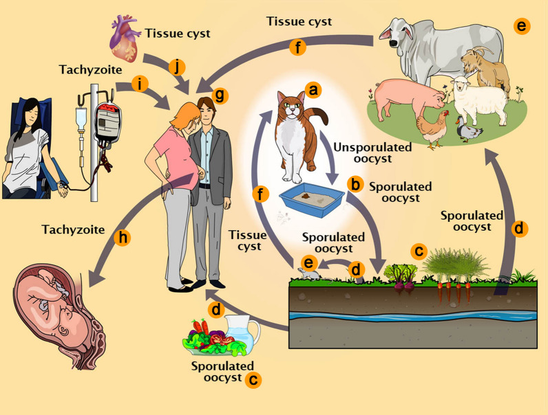
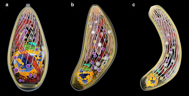
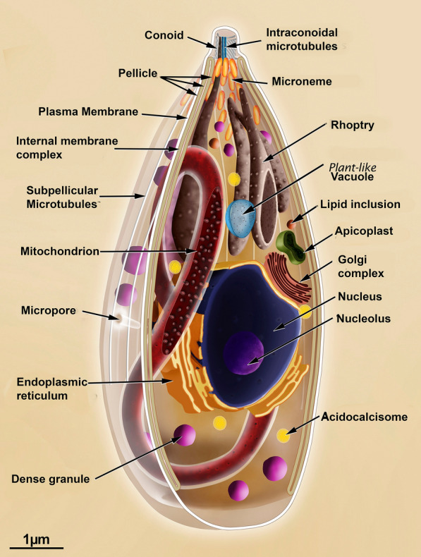
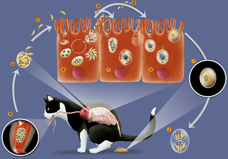
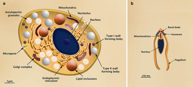
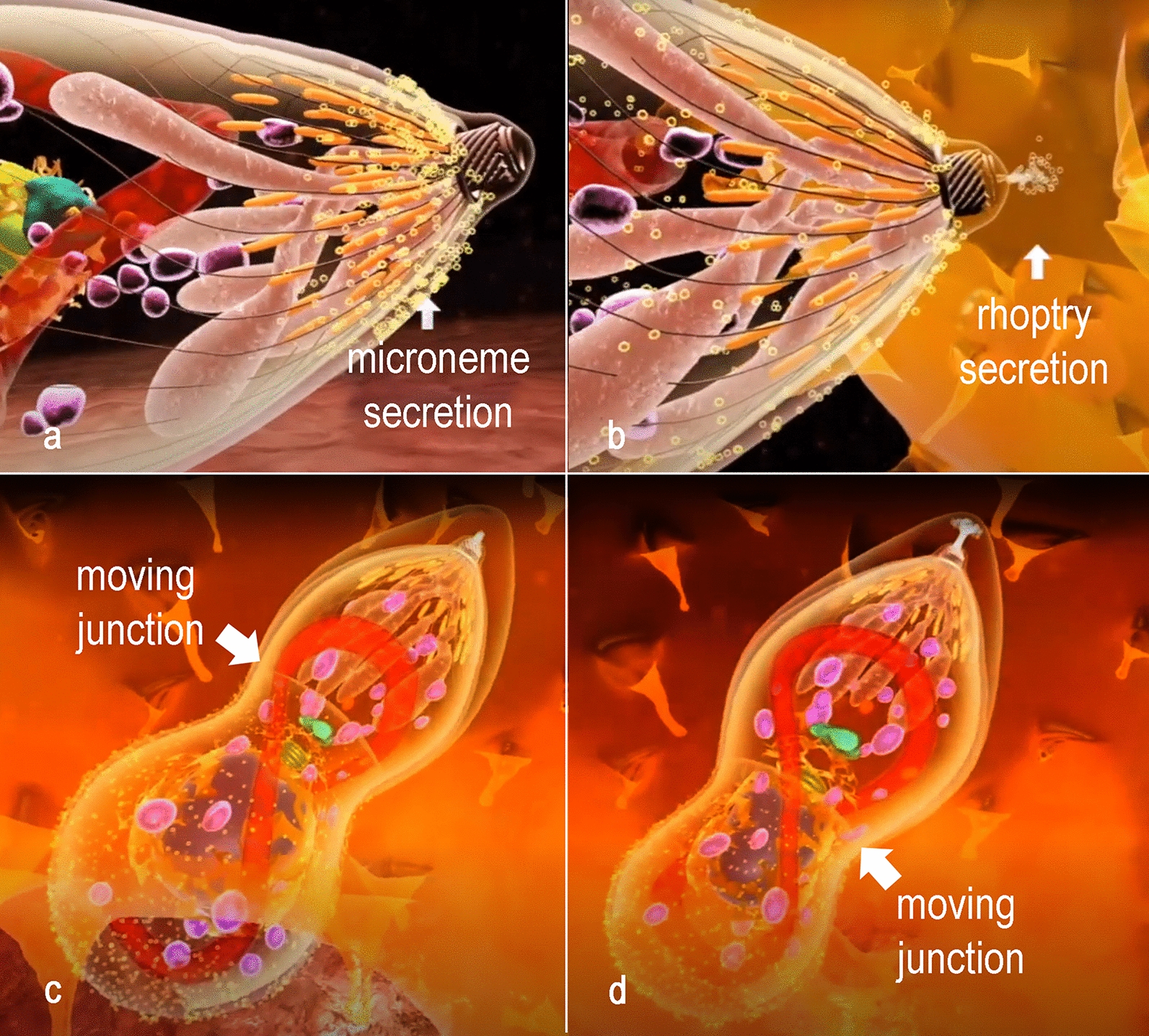
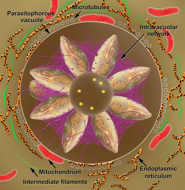
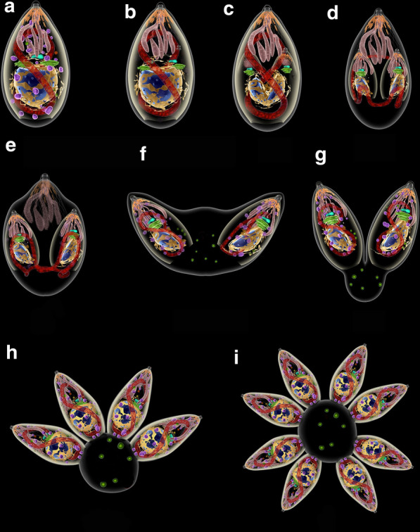
Toxoplasma gondii is a protozoan parasite that is the causative agent of toxoplasmosis, an infection with high prevalence worldwide. Most of the infected individuals are either asymptomatic or have mild symptoms, but T. gondii can cause severe neurologic damage and even death of the fetus when acquired during pregnancy. It is also a serious condition in immunodeficient patients. The life-cycle of T. gondii is complex, with more than one infective form and several transmission pathways. In two animated videos, we describe the main aspects of this cycle, raising questions about poorly or unknown issues of T. gondii biology. Original plates, based on electron microscope observations, are also available for teachers, students and researchers. The main goal of this review is to provide a source of learning on the fundamental aspects of T. gondii biology to students and teachers contributing for better knowledge and control on this important parasite, and unique cell model. In addition, drawings and videos point to still unclear aspects of T. gondii lytic cycle that may stimulate further studies.

Keywords: Apicomplexa; Cell biology; Life-cycle; Parasite; Parasitology; Protozoology; Toxoplasmosis.


Conflict of interest statement

The authors declare that they have no competing interests.

Figures

Fig. 1
Fig. 1
Toxoplasma gondii pathways of transmission. a Feline definitive host (cat). b Unsporulated oocysts in cat feces. c Food contaminated with sporulated oocysts. d Oocysts may be ingested by intermediate hosts via water or raw vegetables. e Intermediate hosts (e.g. cattle, sheep, poultry and swine). f Ingestion of tissue cysts in uncooked meat. g Intermediate hosts (humans). h Tachyzoites transmitted through the placenta to the foetus. i Transmission by blood transfusion and organ transplant (j)
Fig. 2
Fig. 2
The three infective stages of T. gondii. Tachyzoite (a), bradyzoite (b), and sporozoite (c). The nucleus (blue) is surrounded by the rough endoplasmic reticulum (yellow). Above it, The Golgi complex (green) and the apicoplast (blue-green). The single mitochondrion spreads through the cytosol (red). Dense granules (magenta) and amylopectin granules (white) are dispersed in the cytosol. The apical complex is composed by the cylindrical conoid. Below, the secretory organelles: micronemes (orange) and rhoptries (pink). The cell body is limited by three membrane units (the pellicle) and below it a set of subpelicular microtubules
Fig. 3
Fig. 3
Longitudinal section view of the tachyzoite form of Toxoplasma gondii indicating the main structures and organelles
Fig. 4
Fig. 4
Life-cycle of Toxoplasma gondii in cat. a Ingestion of prey containing tissue cysts. b The cyst wall is digested in the stomach and intestines, liberating bradyzoites. c Bradyzoites invade epitelial cells of the intestine. d In the enterocytes bradyzoites divide by schizogony giving rise to merozoites. e Merozoites differentiate into microgamonts, or macrogametes (f). g Fertilization gives rise to an unsporulated oocyst excreted with cat feces (h). i Sporulation occurs and generates two sporocysts with four sporozoites each (j)
Fig. 5
Fig. 5
a Illustration of the macrogamete of Toxoplasma gondii in transversal section showing the main internal structures. b Illustration of the microgamete of Toxoplasma gondii in longitudinal section
Fig. 6
Fig. 6
Sequential events of invasion of a host cell. a Microneme secretion and conoid extrusion are part of the gliding machinery. b The parasite attaches to the host cell membrane and secretion of proteins of the rhoptries neck induces host cell deformation. c, d A moving junction is established between the host cell and the parasite membrane at the point of invasion. Progressively the parasite invades the host cell protected in the PV, from which host cell proteins are excluded, as well as proteins of the gliding machinery
Fig. 7
Fig. 7
Schematic view of a PV in cross section showing tachyzoites linked to the residual body. Inside the residual body, acidocalcisomes (yellow) are accumulated. An intravacuolar network (magenta) stabilizes the rosette of parasites. Profiles of the endoplasmic reticulum (orange) with ribosomes (black) adhered, microtubules (grey circles), intermediate filaments (green) and mitochondria (red) of the host cell and assemble around the PV
Fig. 8
Fig. 8
Sequence of events of division by endodiogeny. a, b The Golgi complex and the apicoplast are the first organelles to divide. c The nucleus assumes a horse-shoe shape. Two new apical complexes start to form. d The inner pellicle grows and embraces the structures of the daughter cells, including the nucleus. e The mitochondrion is the last organelle to be separated between the daughter cells. The apical complex of the mother cell is still maintained at this point. f The two daughter cells emerge and the outer membrane of the mother cell is incorporated. The apical complex of the mother cell disappears. g The two daughter cells remain linked to the residual body where acidocalcisomes (green) start to accumulate. h The process is repeated until a rosette of parasites is formed (i)
Fig. 9
Fig. 9
Scheme of a tissue cyst of Toxoplasma gondii. a The cyst wall is thick and filamentous. Each cyst may contain hundreds of bradyzoites. b A zoom view of the tissue cyst. The cyst is surrounded by a membrane and below it a thick cyst wall is deposited. The components of the cyst wall, as well of the cyst matrix, are secreted by the bradyzoites

Similar articles

Cited by

References

    1. Robert-Gangneux F, Dardé ML. Epidemiology of and diagnostic strategies for toxoplasmosis. Clin Microbiol Rev. 2012;25:264–296. doi: 10.1128/CMR.05013-11. - DOI - PMC - PubMed
    1. Hill D, Dubey JP. Toxoplasma gondii: transmission, diagnosis and prevention. Clin Microbiol Infect. 2002;8:634–640. doi: 10.1046/j.1469-0691.2002.00485.x. - DOI - PubMed
    1. Dubey JP, Lindsay DS, Speer CA. Structures of Toxoplasma gondii tachyzoites, bradyzoites, and sporozoites and biology and development of tissue cysts. Clin Microbiol Rev. 1998;11:267–299. doi: 10.1128/CMR.11.2.267. - DOI - PMC - PubMed
    1. CDC. Toxoplasmosis - epidemiology & risk factors. Atlanta: Centers for Disease Control and Prevention; 2018. https://www.cdc.gov/parasites/toxoplasmosis/epi.html. Accessed 06 Aug 2020.
    1. Dubey JP, Lago EG, Gennari SM, Su C, Jones JL. Toxoplasmosis in humans and animals in Brazil: high prevalence, high burden of disease, and epidemiology. Parasitol. 2012;139:1375–1424. doi: 10.1017/S0031182012000765. - DOI - PubMed