. 2021 Jan 1;206(1):132-140.
doi: 10.4049/jimmunol.2000870. Epub 2020 Nov 23.

The Invariant NKT Cell Response Has Differential Signaling Requirements during Antigen-Dependent and Antigen-Independent Activation

Affiliations

The Invariant NKT Cell Response Has Differential Signaling Requirements during Antigen-Dependent and Antigen-Independent Activation

Courtney K Anderson et al. J Immunol. .

Abstract

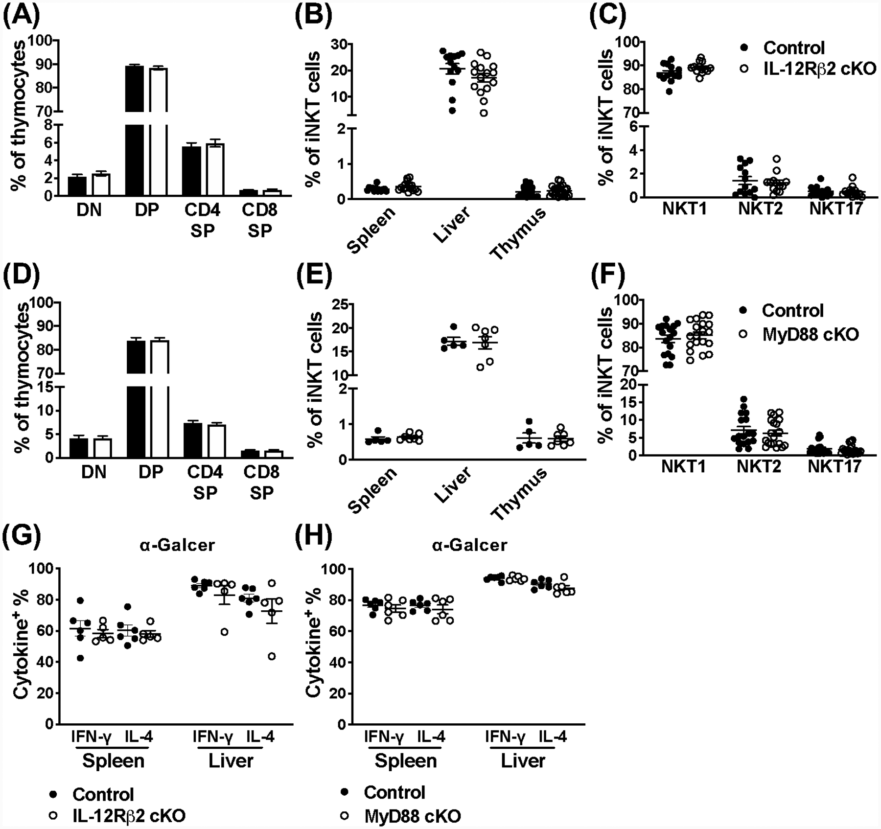
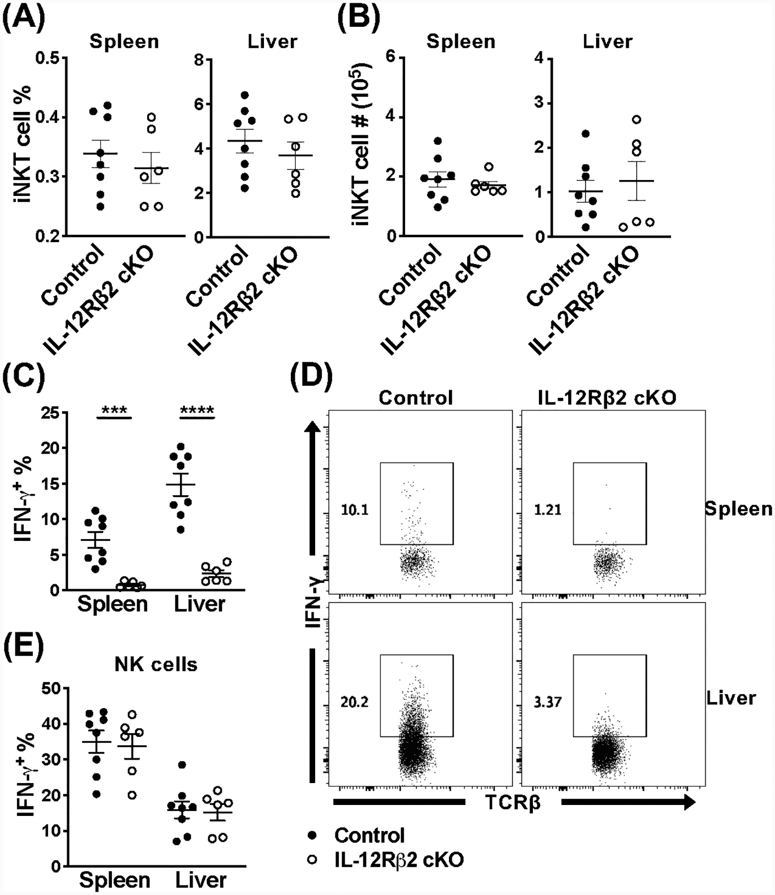
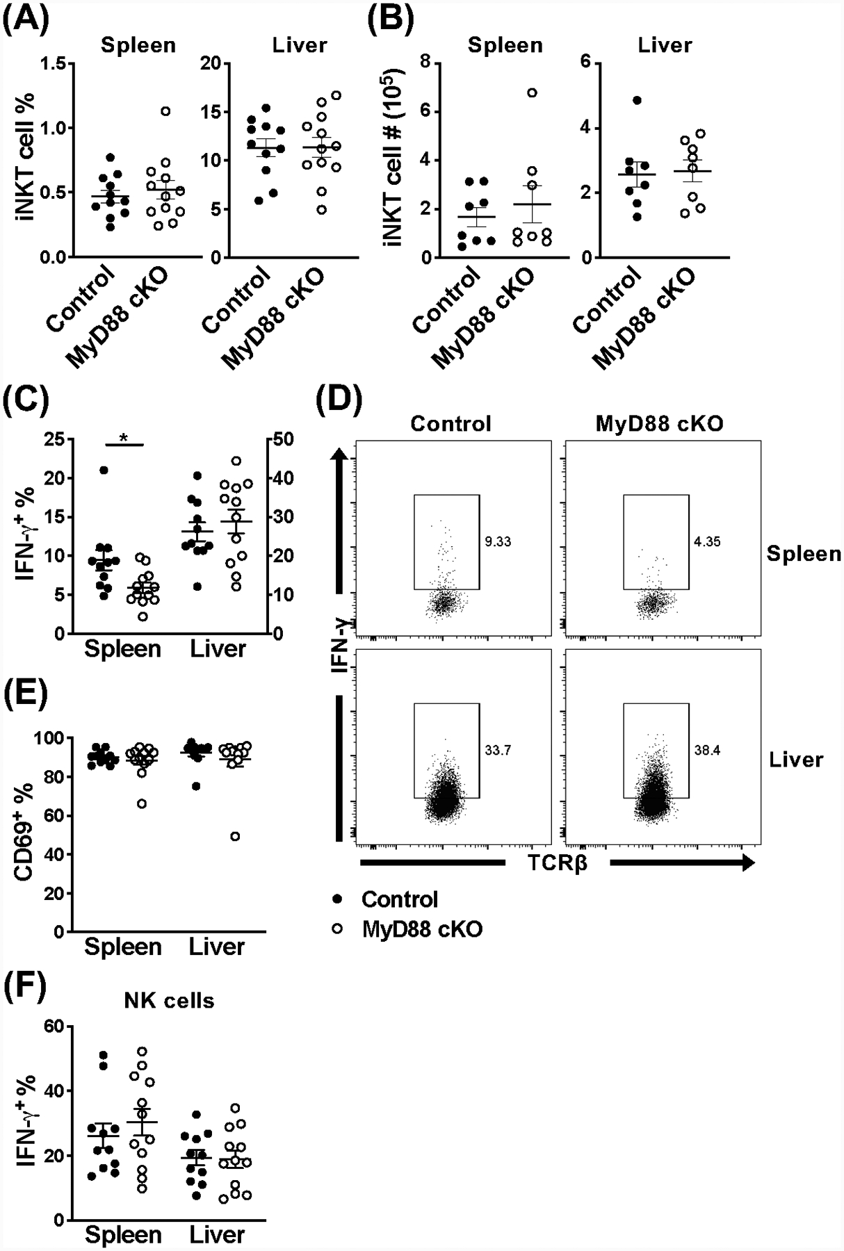
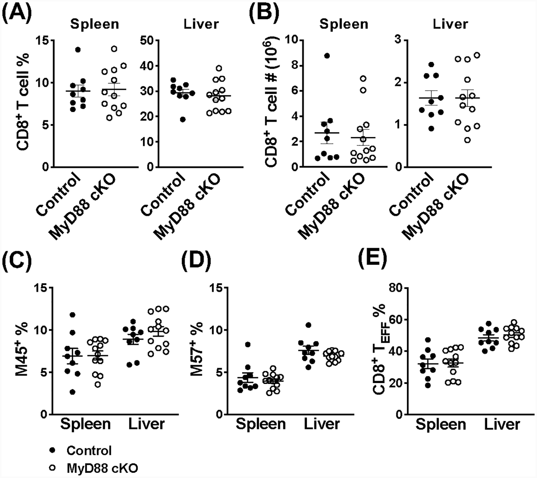
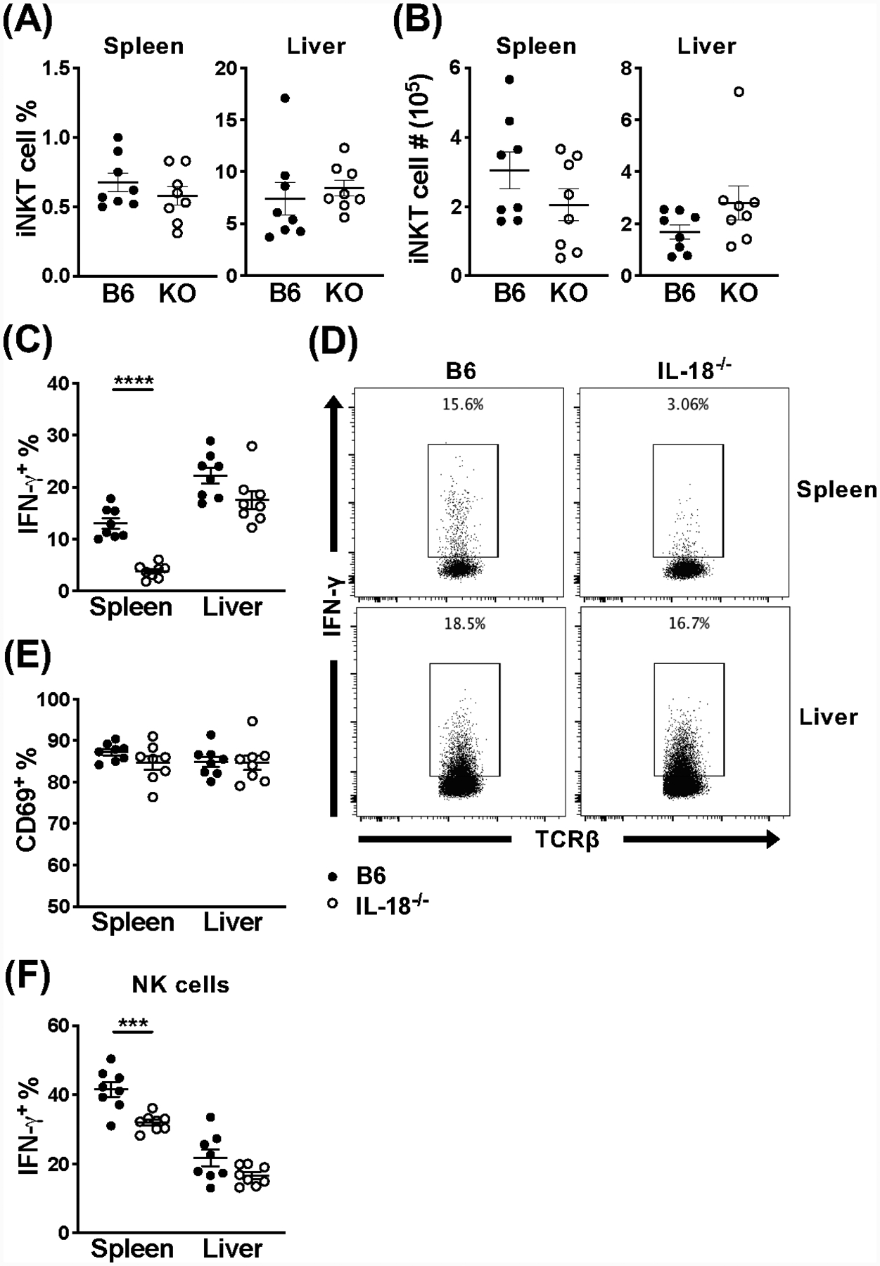
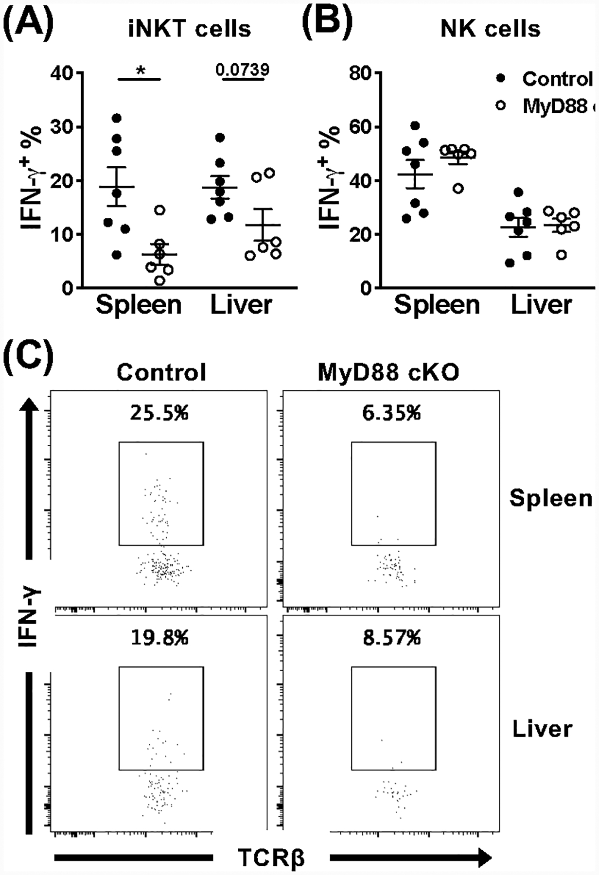
Invariant NKT (iNKT) cells are an innate-like population characterized by their recognition of glycolipid Ags and rapid cytokine production upon activation. Unlike conventional T cells, which require TCR ligation, iNKT cells can also be stimulated independently of their TCR. This feature allows iNKT cells to respond even in the absence of glycolipid Ags, for example, during viral infections. Although the TCR-dependent and -independent activation of iNKT cells have been relatively well established, the exact contributions of IL-12, IL-18, and TLRs remain unclear for these two activation pathways. To definitively investigate how these components affect the direct and indirect stimulation of iNKT cells, we used mice deficient for either MyD88 or the IL-12Rβ2 in the T cell lineage. Using these tools, we demonstrate that IL-12, IL-18, and TLRs are completely dispensable for the TCR activation pathway when a strong agonist is used. In contrast, during murine CMV infection, when the TCR is not engaged, IL-12 signaling is essential, and TLR signaling is expendable. Importantly, to our knowledge, we discovered an intrinsic requirement for IL-18 signaling by splenic iNKT cells but not liver iNKT cells, suggesting that there might be diversity, even within the NKT1 population.


Figures

Figure 1.
Figure 1.
IL-12Rβ2 and MyD88 signaling are dispensable for iNKT cell development, peripheral localization, and their response to a strong agonist. (A) The CD4CD8 double negative (DN), CD4+CD8+ double positive (DP), and CD4+ or CD8 + single positive (SP) stages of T cell development in the thymus of IL-12Rβ2 control (black) and cKO (open) mice (n=18–20). (B) Frequency of iNKT cells (TCRβ+CD1dtet+) in indicated organs from IL-12Rβ2 control (black) and cKO (open) mice (n=13–20). (C) Frequency of NKT1, NKT2, and NKT17 lineages from the thymus of IL-12Rβ2 control (black) and cKO (open) mice (n=13). NKT cell lineages (TCRβ+CD1dtet+) were differentiated using PLZF and RORγt expression. (D) The DN, DP, and CD4+ or CD8+ SP stages of T cell development in the thymus of MyD88 control (black) and cKO (open) mice (n=17). (E) Frequency of iNKT cells (CD45+TCRβ+CD1dtet+) in indicated organs from MyD88 control (black) and cKO (open) mice (n=5–7). (F) Frequency of NKT1, NKT2, and NKT17 lineages from the thymus of MyD88 control (black) and cKO (open) mice (n=17–19). NKT cell lineages (TCRβ+CD1dtet+) were differentiated using PLZF and RORγt expression. (G) Frequency of IFN-γ+ and IL-4+ iNKT cells (TCRβ+CD1dtet+) from the spleen and liver of IL-12Rβ2 control (black) and IL-12Rβ2 cKO (open) mice 2 hours post-stimulation with α-Galcer (n=5–6). (H) Frequency of IFN-γ+ and IL-4+ iNKT cells (TCRβ+CD1dtet+eYFP+) from the spleen and liver of MyD88 control (black) and MyD88 cKO (open) mice 2 hours post-stimulation with α-Galcer (n=6). Data are pooled from two (E, G, H) or at least three (A-D, F) independent experiments and error bars indicate SEM.
Figure 2.
Figure 2.
Loss of IL-12Rβ2 signaling has major effects on iNKT cell activation during acute MCMV infection. (A) Frequency and (B) absolute number of iNKT cells (TCRβ+CD1dtet+) from the spleen and liver of IL-12Rβ2 control (black) and IL-12Rβ2 cKO (open) mice at 36 hours post-infection with MCMV. (C) Frequency and (D) representative flow cytometry of IFN-γ+ iNKT cells (TCRβ+CD1dtet+) and (E) frequency of IFN-γ+ NK cells (TCRβNK1.1+) from the spleen and liver of indicated mice at 36 hours post-MCMV. Data are pooled from or representative of two (A-E, n=6–8) independent experiments and error bars indicate SEM.
Figure 3.
Figure 3.
Splenic iNKT cells are hyporesponsive during MCMV infection in the absence of MyD88 signaling. (A) Frequency and (B) absolute number of iNKT cells (TCRβ+CD1dtet+) from the spleen and liver of MyD88 control (black) and MyD88 cKO (open) mice at 36 hours post-infection with MCMV. (C) Frequency and (D) representative flow cytometry of IFN-γ+ iNKT cells (TCRβ+CD1dtet+eYFP+) and (E) frequency of CD69+ iNKT cells (TCRβ+CD1dtet+eYFP+) from the spleen and liver of indicated mice at 36 hours post-MCMV. (F) Frequency of IFN-γ+ NK cells (TCRβNK1.1+) from the spleen and liver of indicated mice at 36 hours post-MCMV. Data are pooled from two (B, n=8) or three (A, C-F, n=11–12) independent experiments and error bars indicate SEM.
Figure 4.
Figure 4.
CD8+ T cells do not require MyD88 signaling during MCMV infection. (A) Frequency and (B) absolute number of CD8+ T cells (TCRβ+CD8+) from the spleen and liver of MyD88 control (black) and MyD88 cKO (open) mice on day 7 post-infection with MCMV. Frequency of antigen-specific CD8+ T cells (TCRβ+CD8+eYFP+) in the spleen and liver of indicated mice on day 7 post-MCMV using (C) M45-loaded and (D) M57-loaded MHC class I tetramers. (E) Frequency of CD8+ TEFF cells (TCRβ+CD8+eYFP+KLRG1+CD127) from the spleen and liver of indicated mice on day 7 post-MCMV. Data are pooled from three independent experiments (n=9–12) and error bars indicate SEM.
Figure 5.
Figure 5.
NKT1 cells are not detrimentally affected by loss of MyD88 signaling during acute MCMV infection. (A) Frequency and (B) absolute number of NKT1, NKT2, and NKT17 cells (CD45+CD1dtet+TCRβ+) in the spleen and liver of MyD88 control (black) and MyD88 cKO (open) animals at 36 hours post-infection with MCMV. NKT cell lineages were differentiated using PLZF, RORγt, and T-bet expression. Data are pooled from two independent experiments (n=9–10) and error bars indicate SEM.
Figure 6.
Figure 6.
Loss of IL-18 signaling results in hyporesponsive splenic iNKT cells, however hepatic iNKT cells respond normally. (A) Frequency and (B) absolute number of iNKT cells (CD45+TCRβ+CD1dtet+) from the spleen and liver of C57BL/6 (black) or IL-18−/− (open) mice at 36 hours post-infection with MCMV. (C) Frequency and (D) representative flow cytometry of IFN-γ+ iNKT cells (CD45+TCRβ+CD1dtet+) and (E) frequency of CD69+ iNKT cells (CD45+TCRβ+CD1dtet+) from the spleen and liver of indicated mice at 36 hours post-MCMV. (F) Frequency of IFN-γ+ NK cells (CD45+TCRβNK1.1+) from the spleen and liver of indicated mice at 36 hours post-MCMV. Data are pooled from or representative of two independent experiments (n=8) and error bars indicate SEM.
Figure 7.
Figure 7.
Splenic iNKT cells are intrinsically affected by loss of MyD88 signaling during acute MCMV infection. Frequency of IFN-γ+ donor (A) iNKT cells (CD45+TCRβ+CD1dtet+CD45.1CD45.2+eYFP+) and (B) NK cells (CD45+TCRβNK1.1+CD45.1CD45.2+) from the spleen and liver of B6.SJL recipients at 36 hours post-infection with MCMV. Donor cells were isolated from either MyD88 control (black) or MyD88 cKO (open) animals and enriched for iNKT cells prior to adoptive transfer. (C) Representative flow cytometry of IFN-γ production by indicated donor iNKT cells from the spleen and liver of B6.SJL recipients. Data are pooled from or representative of two independent experiments (n=6–7) and error bars indicate SEM.

References

    1. Beckman EM, Porcelli SA, Morita CT, Behar SM, Furlong ST, and Brenner MB. 1994. Recognition of a lipid antigen by CD1-restricted alpha beta+ T cells. Nature 372: 691–694. - PubMed
    1. Crowe NY, Uldrich AP, Kyparissoudis K, Hammond KJ, Hayakawa Y, Sidobre S, Keating R, Kronenberg M, Smyth MJ, and Godfrey DI. 2003. Glycolipid antigen drives rapid expansion and sustained cytokine production by NK T cells. Journal of immunology (Baltimore, Md. : 1950) 171: 4020–4027. - PubMed
    1. Wilson MT, Johansson C, Olivares-Villagomez D, Singh AK, Stanic AK, Wang CR, Joyce S, Wick MJ, and Van Kaer L. 2003. The response of natural killer T cells to glycolipid antigens is characterized by surface receptor down-modulation and expansion. Proceedings of the National Academy of Sciences of the United States of America 100: 10913–10918. - PMC - PubMed
    1. Matsuda JL, Gapin L, Baron JL, Sidobre S, Stetson DB, Mohrs M, Locksley RM, and Kronenberg M. 2003. Mouse V alpha 14i natural killer T cells are resistant to cytokine polarization in vivo. Proceedings of the National Academy of Sciences of the United States of America 100: 8395–8400. - PMC - PubMed
    1. Stetson DB, Mohrs M, Reinhardt RL, Baron JL, Wang ZE, Gapin L, Kronenberg M, and Locksley RM. 2003. Constitutive cytokine mRNAs mark natural killer (NK) and NK T cells poised for rapid effector function. The Journal of experimental medicine 198: 1069–1076. - PMC - PubMed

Publication types