. 2020 Dec 8;117(49):31417-31426.
doi: 10.1073/pnas.2013237117. Epub 2020 Nov 23.

Studies in bank voles reveal strain differences between chronic wasting disease prions from Norway and North America

Affiliations

Studies in bank voles reveal strain differences between chronic wasting disease prions from Norway and North America

Romolo Nonno et al. Proc Natl Acad Sci U S A. .

Abstract

Chronic wasting disease (CWD) is a relentless epidemic disorder caused by infectious prions that threatens the survival of cervid populations and raises increasing public health concerns in North America. In Europe, CWD was detected for the first time in wild Norwegian reindeer (Rangifer tarandus) and moose (Alces alces) in 2016. In this study, we aimed at comparing the strain properties of CWD prions derived from different cervid species in Norway and North America. Using a classical strain typing approach involving transmission and adaptation to bank voles (Myodes glareolus), we found that prions causing CWD in Norway induced incubation times, neuropathology, regional deposition of misfolded prion protein aggregates in the brain, and size of their protease-resistant core, different from those that characterize North American CWD. These findings show that CWD prion strains affecting Norwegian cervids are distinct from those found in North America, implying that the highly contagious North American CWD prions are not the proximate cause of the newly discovered Norwegian CWD cases. In addition, Norwegian CWD isolates showed an unexpected strain variability, with reindeer and moose being caused by different CWD strains. Our findings shed light on the origin of emergent European CWD, have significant implications for understanding the nature and the ecology of CWD in Europe, and highlight the need to assess the zoonotic potential of the new CWD strains detected in Europe.

Keywords: CWD; TSE; chronic wasting disease; prion strains; prions.


Conflict of interest statement

The authors declare no competing interest.

Figures
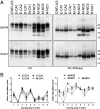
Fig. 1.
Fig. 1.
PrPSc types and neurodegeneration in Bv109I infected with CWD isolates. (A) Representative replica Western blots showing PK-treated (PK) or PK-treated and deglycosylated (PK+PNGase) PrPSc from the brains of voles inoculated with the CWD isolates indicated on the Top of the blots, and analyzed with mAbs recognizing different epitopes on PrP, as indicated on the Left of the blots (SAF84 epitope at 167 to 173; Sha31 epitope at 146 to 153). The CWD inocula are indicated as in Table 1; CWD-US in the first lane indicates a Bv109I inoculated in a previous study (24) with an elk CWD isolate from the United States, here analyzed for comparison purposes. The positions of MW markers are indicated on the Right of the blots (in kilodaltons). Note that a PrPres fragment migrating at ∼12 to 13 kDa was detected by SAF84 only in Bv109I inoculated with Norwegian moose isolates. This PrPres fragment was C-terminal and N-glycosylated, as it was not recognized by the more N-terminal Sha31 mAb and was enriched upon removal of N-linked sugars. (B) Lesion profiles in groups of Bv109I infected with CWD isolates from Canadian cervids (Left) or Norwegian moose (Right). Data points represent the mean ± SEM of at least five voles per group. Brain-scoring areas: medulla (1), cerebellum (2), superior colliculus (3), hypothalamus (4), thalamus (5), hippocampus (6), septum (7), retrosplenial and adjacent motor cortex (8), and cingulate and adjacent motor cortex (9).
Fig. 2.
Fig. 2.
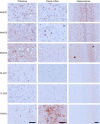
Immunohistochemical patterns of PrPSc deposition in Bv109I inoculated with CWD isolates. Immunohistochemical detection of PrPSc in the thalamus, visual cortex (third layer), and hippocampus of Bv109I inoculated with the CWD isolates indicated on the Left. Norwegian moose isolates (M-NO1, M-NO2, and M-NO3) were characterized by strong PrPSc deposition in all brain areas, which was mainly intraneuronal, particularly in the case of M-NO1 and M-NO2. In contrast, PrPSc deposition was milder in Bv109I inoculated with Canadian isolates M-CA1 and E-CA2 and was mainly detected as diffuse deposits in the thalamus. The only Bv109I that was positive after inoculation of R-NO1 was characterized by a strong and diffuse PrPSc deposition in the visual cortex. PrPSc was detected by mAb SAF84. (Scale bar: 50 µm.)
Fig. 3.
Fig. 3.
PrPSc types and neurodegeneration in Bv109I-adapted CWD isolates. (A) Representative replica Western blots showing PK-treated PrPSc from Bv109I after second or third subpassages with the CWD isolates indicated on the Top of the blots, and analyzed with mAbs recognizing different epitopes on PrP, as indicated below the blots (SAF84 epitope at 167 to 173; Sha31 epitope at 146 to 153; 12B2 epitope at 93 to 97). The positions of molecular weight markers are indicated on the Right of the blots (in kilodaltons). Note that the PrPres fragment migrating at ∼12 to 13 kDa was detected by SAF84 only in Bv109I after second and third passages with Norwegian moose isolates and was much more abundant in M-NO2 than in M-NO1. Furthermore, in M-NO2 there was a decrease of the main PrPres fragment at the third passage (Sha31 blot), which was also reflected in a decreased 12B2 binding. The blot detected with Sha31 shows that PrPres in M-NO1 (second and third passages) and R-NO1 migrates slightly higher than in E-CA1, E-CA2, D-CA1, M-CA1, and M-NO2 (second and third passages). This was reflected in a better preservation of the N-terminal 12B2 epitope as evidenced by the partial loss of PrPres signal in E-CA1, E-CA2, D-CA1, M-CA1, and M-NO2, but not in M-NO1 and R-NO1, in the blot detected with 12B2. (B) Lesion profiles in groups of Bv109I after two passages with CWD isolates from Norwegian moose (Upper), Canadian cervids (Middle), or Norwegian reindeer (Lower). Data points represent the mean (SEM) of at least five voles per group. Brain-scoring areas: medulla (1), cerebellum (2), superior colliculus (3), hypothalamus (4), thalamus (5), hippocampus (6), septum (7), retrosplenial and adjacent motor cortex (8), and cingulate and adjacent motor cortex (9).
Fig. 4.
Fig. 4.
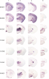
Different patterns of PrPSc deposition in Bv109I-adapted CWD isolates. PET blot detection of protease-resistant PrPSc in coronal sections of the forebrain, representing telencephalon (1), diencephalon (2), midbrain (3), and hindbrain (4) from representative Bv109I following two passages with the CWD isolates indicated on the Left. PrPSc was detected with mAb SAF84.
Fig. 5.
Fig. 5.
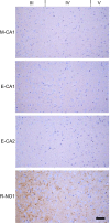
Different patterns of PrPSc deposition in cortical layers of Bv109I-adapted CWD isolates. Immunohistochemical detection of PrPSc in the third, fourth, and fifth cortical layers of the somatosensory cortex of Bv109I after second passage with the CWD isolates indicated on the Left. PrPSc was not detected in Bv109I infected with the Canadian CWD isolates but was found as diffuse and punctate deposits in all layers of Bv109I infected with R-NO1. PrPSc was detected by mAb SAF84. (Scale bar: 50 µm.)
Fig. 6.
Fig. 6.
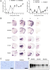
Different neuropathological and PrPSc phenotypes induced by Norwegian and Canadian CWD isolates after three passages in Bv109I. Comparison of neuropathological and molecular phenotypes for groups of Bv109I analyzed by different techniques shows four different phenotypes of disease in Bv109I-adapted CWD from Canadian isolates, M-NO1, M-NO2, and R-NO1, respectively. (A) Lesion profiles in Bv109I following the third passages of CWD isolates from Norwegian moose (M-NO1 and M-NO2, Left), Canadian CWD isolates and Norwegian reindeer (E-CA1, M-CA1, and R-NO1, Right). Data points represent the mean (SEM) of at least five voles per group. Brain-scoring areas: medulla (1), cerebellum (2), superior colliculus (3), hypothalamus (4), thalamus (5), hippocampus (6), septum (7), retrosplenial and adjacent motor cortex (8), and cingulate and adjacent motor cortex (9). (B) PET blot detection of protease-resistant PrPSc in brain coronal sections representing telencephalon (1), diencephalon (2), midbrain (3), and hindbrain (4) from representative Bv109I after the third passages with the CWD isolates M-NO1, M-NO2, M-CA1, E-CA1, and R-NO1, as indicated. Note that, in agreement with the absence of spongiform degeneration in the medulla of Bv109I infected with M-NO2 but not with M-NO1 (see A), PrPSc deposition in the hindbrain was low or absent in M-NO2. M-CA1 and E-CA1 showed a similar pattern of PrPSc deposition, which was different from M-NO1 and M-NO2, in agreement with the different lesion profiles (see A). PrPSc deposition in R-NO1 mostly overlapped with that in M-CA1 and E-CA1, but was characterized by the involvement of superficial cortical layers in the somatosensory cortex, which was absent in Bv109I infected with Canadian CWD isolates (arrowheads in coronal sections A and B). PrPSc was detected with mAb SAF84. (C) Immunohistochemical detection of PrPSc in the corpus callosum of Bv109I inoculated with the CWD isolates M-NO1 and M-NO2, showing PrPSc deposition in Bv109I infected with M-NO2 but not in those with M-NO1. PrP was detected by mAb SAF84. (Scale bar: 50 µm.) (D) Western blot showing PK-treated PrPSc from three Bv109I per group after second passages with R-NO1, E-CA1, and M-CA1, as indicated on the Top of the blot. PrPSc was detected with mAb SAF84. The position of the MW markers is indicated on the Right of the blots (in kilodaltons).

References

    1. Ironside J. W., Ritchie D. L., Head M. W., Prion diseases. Handb. Clin. Neurol. 145, 393–403 (2017). - PubMed
    1. Knight R., Infectious and sporadic prion diseases. Prog. Mol. Biol. Transl. Sci. 150, 293–318 (2017). - PubMed
    1. Houston F., Andréoletti O., Animal prion diseases: The risks to human health. Brain Pathol. 29, 248–262 (2019). - PMC - PubMed
    1. Rivera N. A., Brandt A. L., Novakofski J. E., Mateus-Pinilla N. E., Chronic wasting disease in cervids: Prevalence, impact and management strategies. Vet. Med. (Auckl.) 10, 123–139 (2019). - PMC - PubMed
    1. Kim T. Y., et al., Additional cases of chronic wasting disease in imported deer in Korea. J. Vet. Med. Sci. 67, 753–759 (2005). - PubMed

Publication types

LinkOut - more resources