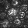
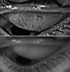
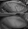
A review of meibography for a refractive surgeon
- PMID: 33229641
- PMCID: PMC7856926
- DOI: 10.4103/ijo.IJO_2465_20
A review of meibography for a refractive surgeon
Abstract
Refractive surgery has evolved from being a therapeutic correction of high refractive errors to a cosmetic correction. The expectations associated with such a surgery are enormous and one has to anticipate all possible complications and side-effects that come with the procedure and prepare accordingly. The most common amongst these is post-refractive surgery dry eye of which Meibomian gland dysfunction is a commonly associated cause. We present an understanding of various diagnostic imaging modalities that can be used for evaluating meibomian glands which can also serve as a visual aid for patient understanding. We also describe various common conditions which can silently cause changes in the gland architecture and function which are to be considered and evaluated for.
Keywords: Dry eye disease; meibography; meibomian gland dysfunction.
Conflict of interest statement
None
Figures


References
-
- Lekhanont K, Rojanaporn D, Chuck RS, Vongthongsri A. Prevalence of dry eye in Bangkok, Thailand. Cornea. 2006;25:1162–7. - PubMed
-
- Jie Y, Xu L, Wu YY, Jonas JB. Prevalence of dry eye among adult Chinese in the Beijing Eye Study. Eye (Lond) 2009;23:688–93. - PubMed
-
- Basak SK, Pal PP, Basak S, Bandyopadhyay A, Choudhury S, Sar S. Prevalence of dry eye diseases in hospital-based population in West Bengal, Eastern India. J Indian Med Assoc. 2012;110:789–94. - PubMed
-
- Chatterjee S, Agrawal D, Sharma A. Meibomian gland dysfunction in a hospital-based population in central India. Cornea. 2020;39:634–9. - PubMed
Publication types
MeSH terms
LinkOut - more resources
Full Text Sources
Medical

